This book deals with the quantitative (mensural) aspects of Electron Micrography (EM) [obtained by using both, Transmission (TEM) and Scanning (SEM) types] by applying Photogrammetric techniques.
This unique book on super-resolution microscopy techniques presents comparative, in-depth analyses of the strengths and weaknesses of the individual approaches.
This unique book on super-resolution microscopy techniques presents comparative, in-depth analyses of the strengths and weaknesses of the individual approaches.
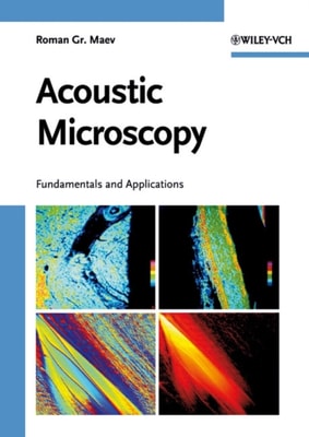
Novel physical solutions, including new results in the field of adaptive methods and inventive approaches to inverse problems, original concepts based on high harmonic imaging algorithms, intriguing vibro-acoustic imaging and vibro-modulation technique, etc.
Novel physical solutions, including new results in the field of adaptive methods and inventive approaches to inverse problems, original concepts based on high harmonic imaging algorithms, intriguing vibro-acoustic imaging and vibro-modulation technique, etc.
Adopting a didactical approach from fundamentals to actual experiments and applications, this handbook and ready reference covers real-time observations using modern scanning electron microscopy and transmission electron microscopy, while also providing information on the required stages and samples.
Adopting a didactical approach from fundamentals to actual experiments and applications, this handbook and ready reference covers real-time observations using modern scanning electron microscopy and transmission electron microscopy, while also providing information on the required stages and samples.
About 40 % of current atomic force microscopy (AFM) research is performed in liquids, making liquid-based AFM a rapidly growing and important tool for the study of biological materials.
About 40 % of current atomic force microscopy (AFM) research is performed in liquids, making liquid-based AFM a rapidly growing and important tool for the study of biological materials.
Filling a gap in the literature, this book features in-depth discussions on amplitude modulation AFM, providing an overview of the theory, instrumental considerations and applications of the technique in both academia and industry.
Well-structured and adopting a pedagogical approach, this self-contained monograph covers the fundamentals of scanning probe microscopy, showing how to use the techniques for investigating physical and chemical properties on the nanoscale and how they can be used for a wide range of soft materials.
Well-structured and adopting a pedagogical approach, this self-contained monograph covers the fundamentals of scanning probe microscopy, showing how to use the techniques for investigating physical and chemical properties on the nanoscale and how they can be used for a wide range of soft materials.
Filling a gap in the literature, this book features in-depth discussions on amplitude modulation AFM, providing an overview of the theory, instrumental considerations and applications of the technique in both academia and industry.
This only and up-to-date monograph on this versatile method covers its use in a range of applications spanning the fields of physics, materials science, electrical engineering, medicine, and research and industry.
Derived from the successful three-volume Handbook of Microscopy, this book provides a broad survey of the physical fundamentals and principles of all modern techniques of electron microscopy.
This new and completely updated edition features not only an accompanying CD-ROM, but also a new applications section, reflecting the many breakthroughs in the field over the last few years.
This first book to focus on the use of SPMs to actively manipulate molecules and nanostructures on surfaces goes way beyond conventional treatments of scanning microscopy merely for imaging purposes.
"e;When you first view Rose-Lynn Fisher's photographs, you might think you're looking down at the world from an airplane, at dunes, skyscrapers or shorelines.
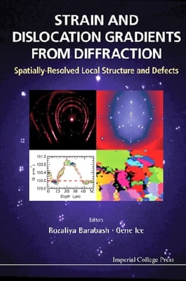
This book highlights emerging diffraction studies of strain and dislocation gradients with mesoscale resolution, which is currently a focus of research at laboratories around the world.
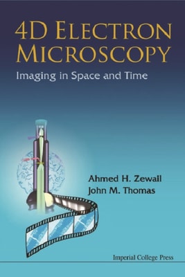
The modern electron microscope, as a result of recent revolutionary developments and many evolutionary ones, now yields a wealth of quantitative knowledge pertaining to structure, dynamics, and function barely matched by any other single scientific instrument.
Atomic force microscopy (AFM) is part of a range of emerging microscopic methods for biologists which offer the magnification range of both the light and electron microscope, but allow imaging under the 'natural' conditions usually associated with the light microscope.
Atomic Force Microscopy: Principles, Developments and Applications presents Atomic Force Microscopy (AFM) as one of the most powerful tools for the analysis of morphologies because it creates three-dimensional images at the angstrom and nano scale.
Microscopy is a dynamic area of science, incorporating both basic classroom microscopes and sophisticated research style instruments that can be driven by light, electrons, or X-rays.
Twenty-two experienced scientists from eleven different countries have contributed four years of study and discussion to this important book, which represents part of the work done by the International Committee on Microbiological Specifications for Foods, a standing committee of the International Association of Microbiological Societies.
Microscopy is a dynamic area of science, incorporating both basic classroom microscopes and sophisticated research style instruments that can be driven by light, electrons, or X-rays.
A guide to modern scanning electron microscopy instrumentation, methodology and techniques, highlighting novel applications to cell and molecular biology.
A guide to modern scanning electron microscopy instrumentation, methodology and techniques, highlighting novel applications to cell and molecular biology.
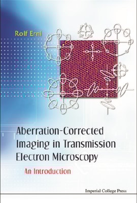
The book is concerned with the theory, background, and practical use of transmission electron microscopes with lens correctors that can correct the effects of spherical aberration.
The book is concerned with the theory, background, and practical use of transmission electron microscopes with lens correctors that can correct the effects of spherical aberration.
DIATOM MICROSCOPY The main goal of the book is to demonstrate the wide variety of microscopy methods being used to investigate natural and altered diatom structures.
DIATOM MICROSCOPY The main goal of the book is to demonstrate the wide variety of microscopy methods being used to investigate natural and altered diatom structures.
A comprehensive guide to the art and science of bioimaging data acquisition, processing and analysis Standard and Super-Resolution Bioimaging Data Analysis gets newcomers to bioimage data analysis quickly up to speed on the mathematics, statistics, computing hardware and acquisition technologies required to correctly process and document data.
A comprehensive guide to the art and science of bioimaging data acquisition, processing and analysis Standard and Super-Resolution Bioimaging Data Analysis gets newcomers to bioimage data analysis quickly up to speed on the mathematics, statistics, computing hardware and acquisition technologies required to correctly process and document data.
Describes new state-of-the-science tools and their contribution to industrial R&D With contributions from leading international experts in the field, this book explains how scanning probe microscopy is used in industry, resulting in improved product formulation, enhanced processes, better quality control and assurance, and new business opportunities.
Describes new state-of-the-science tools and their contribution to industrial R&D With contributions from leading international experts in the field, this book explains how scanning probe microscopy is used in industry, resulting in improved product formulation, enhanced processes, better quality control and assurance, and new business opportunities.
A detailed presentation of the physics of electron beam-specimen interactions Electron microscopy is one of the most widely used characterisation techniques in materials science, physics, chemistry, and the life sciences.
A detailed presentation of the physics of electron beam-specimen interactions Electron microscopy is one of the most widely used characterisation techniques in materials science, physics, chemistry, and the life sciences.
The go to resource for microscopists on biological applications of field emission gun scanning electron microscopy (FEGSEM) The evolution of scanning electron microscopy technologies and capability over the past few years has revolutionized the biological imaging capabilities of the microscope giving it the capability to examine surface structures of cellular membranes to reveal the organization of individual proteins across a membrane bilayer and the arrangement of cell cytoskeleton at a nm scale.
The go to resource for microscopists on biological applications of field emission gun scanning electron microscopy (FEGSEM) The evolution of scanning electron microscopy technologies and capability over the past few years has revolutionized the biological imaging capabilities of the microscope giving it the capability to examine surface structures of cellular membranes to reveal the organization of individual proteins across a membrane bilayer and the arrangement of cell cytoskeleton at a nm scale.
Part of the Wiley-Royal Microscopical Society Series, this book discusses the rapidly developing cutting-edge field of low-voltage microscopy, a field that has only recently emerged due to the rapid developments in the electron optics design and image processing.
Part of the Wiley-Royal Microscopical Society Series, this book discusses the rapidly developing cutting-edge field of low-voltage microscopy, a field that has only recently emerged due to the rapid developments in the electron optics design and image processing.
Fundamentals of Light Microscopy and Electronic Imaging, Second Edition provides a coherent introduction to the principles and applications of the integrated optical microscope system, covering both theoretical and practical considerations.
Fundamentals of Light Microscopy and Electronic Imaging, Second Edition provides a coherent introduction to the principles and applications of the integrated optical microscope system, covering both theoretical and practical considerations.