With every chapter revised and updated, Physics for Diagnostic Radiology, Third Edition continues to emphasise the importance of physics education as a critical component of radiology training.
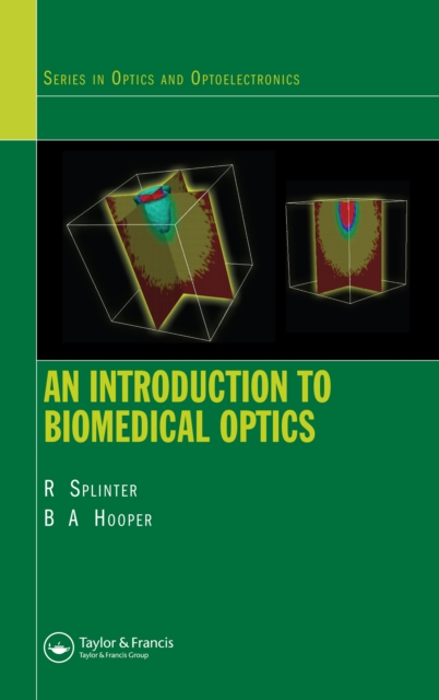
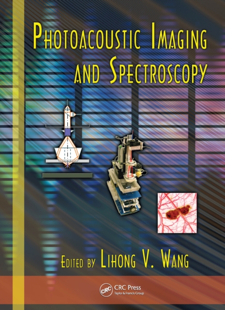
Despite a number of books on biophotonics imaging for medical diagnostics and therapy, the field still lacks a comprehensive imaging book that describes state-of-the-art biophotonics imaging approaches intensively developed in recent years.
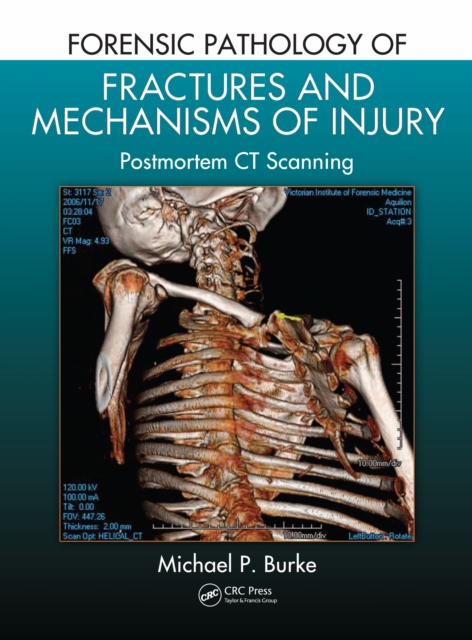
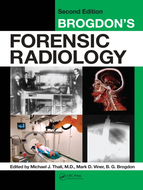
Practitioners of forensic medicine have various tools at their disposal to determine cause of death, and today's computed tomography (CT) can provide valuable clues if images are interpreted properly.
Conventional computed tomography (CT) techniques employ a narrow array of x-ray detectors and a fan-shaped x-ray beam to rotate around the patient to produce images of thin sections of the patient.
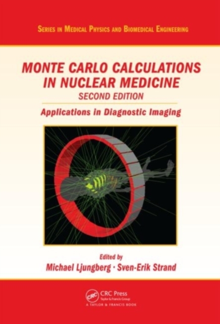
From first principles to current computer applications, Monte Carlo Calculations in Nuclear Medicine, Second Edition: Applications in Diagnostic Imaging covers the applications of Monte Carlo calculations in nuclear medicine and critically reviews them from a diagnostic perspective.
More work is being done in the statistical aspects of medical imaging, and this book fills the gap to provide a unified framework of study by presenting a complete look at medical imaging and statistics - from the statistical aspects of imaging technology to the statistical analysis of images.
Bringing the long tradition of radiologic pathologic correlation to forensic radiology and autopsy, this volume provides readers with a technical and interpretive foundation for applying modern cross-sectional imaging to forensic autopsy.
The benchmark first edition of Forensic Radiology, published in 1998, was a milestone in the forensic community a "e; a bestseller throughout the world and a standard reference for practitioners and educators alike.
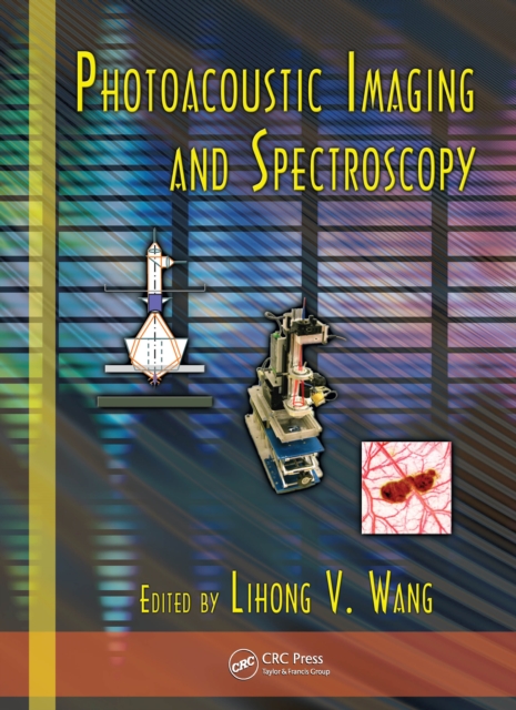
Photoacoustics promises to revolutionize medical imaging and may well make as dramatic a contribution to modern medicine as the discovery of the x-ray itself once did.
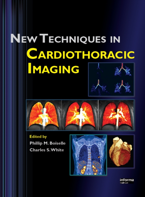
New Techniques in Cardiothoracic Imaging emphasizes emerging methods in computed tomography, magnetic resonance imaging, positron-emission tomography, and similar technology.
Photoacoustics promises to revolutionize medical imaging and may well make as dramatic a contribution to modern medicine as the discovery of the x-ray itself once did.
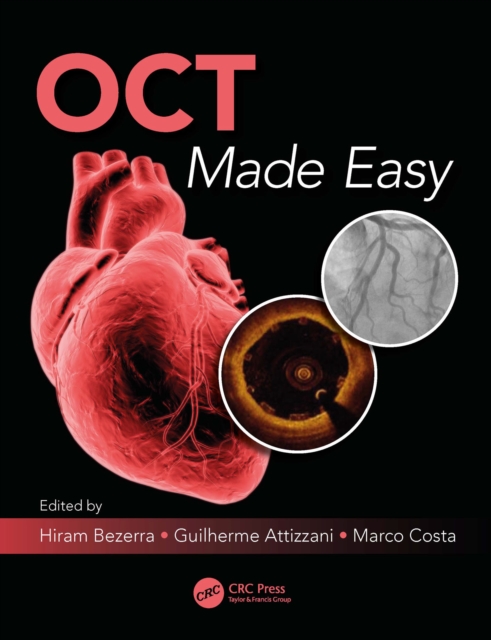
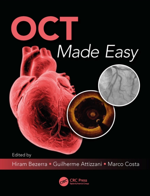
This book, written by premier authors in the field of OCT intravascular imaging, covers the best practices for using OCT to provide high resolution cross-sectional viewing for atherosclerotic plaque assessment, stent strut coverage and apposition, assessment in stent restenosis evaluation, and PCI guide and optimization.
This book, written by premier authors in the field of OCT intravascular imaging, covers the best practices for using OCT to provide high resolution cross-sectional viewing for atherosclerotic plaque assessment, stent strut coverage and apposition, assessment in stent restenosis evaluation, and PCI guide and optimization.
This book describes methods and procedures for preparing PET radiopharmaceuticals, and highlights new methods for conducting radiochemical reactions with carbon-11 (C11) and fluorine-18 (F18), which are two of the most commonly used radionuclides in positron emission tomography (PET) imaging.
This book describes methods and procedures for preparing PET radiopharmaceuticals, and highlights new methods for conducting radiochemical reactions with carbon-11 (C11) and fluorine-18 (F18), which are two of the most commonly used radionuclides in positron emission tomography (PET) imaging.
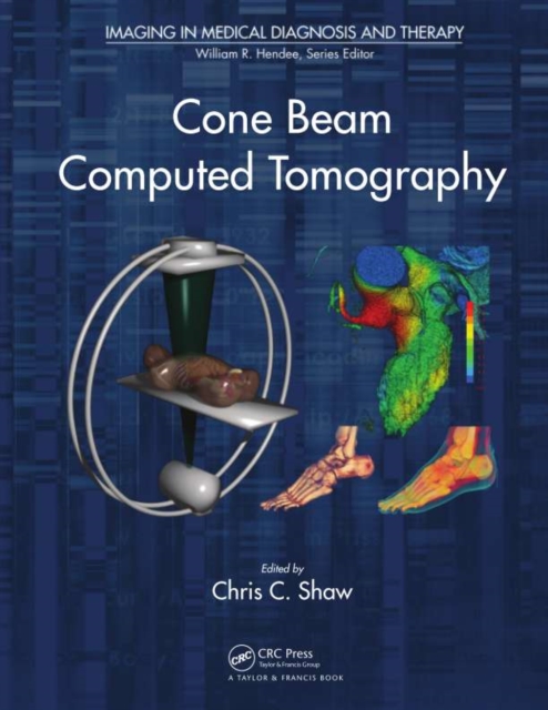
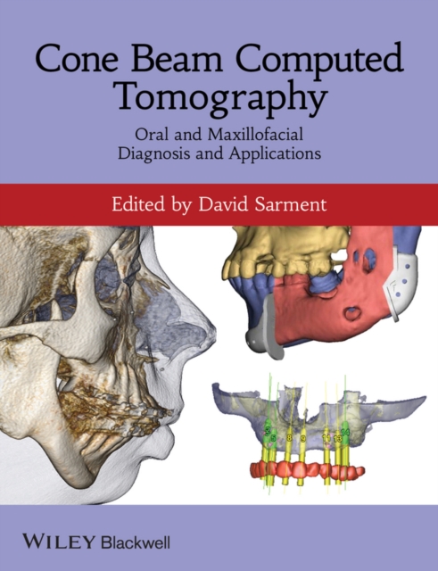
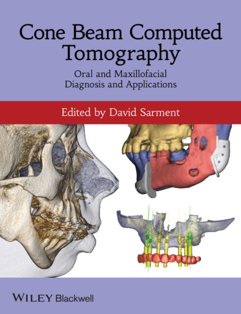
Written for the clinician, Cone Beam Computed Tomography helps the reader understand how CBCT machines operate, perform advanced diagnosis using CT data, have a working knowledge of CBCT-related treatment planning for specific clinical tasks, and integrate these new technologies in daily practice.
Written for the clinician, Cone Beam Computed Tomography helps the reader understand how CBCT machines operate, perform advanced diagnosis using CT data, have a working knowledge of CBCT-related treatment planning for specific clinical tasks, and integrate these new technologies in daily practice.
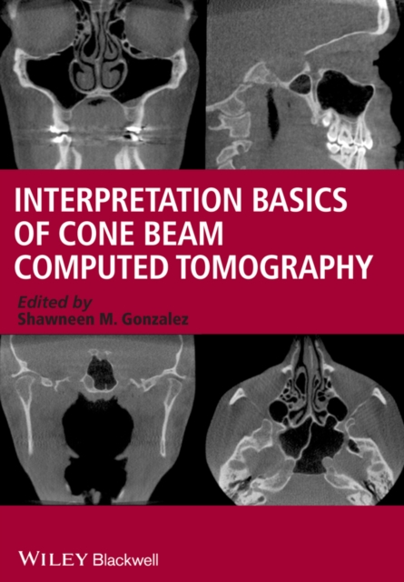
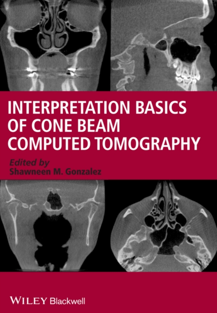
Interpretation Basics of Cone Beam Computed Tomography is an easy-to-use guide to Cone Beam CT technology for general dental practitioners and dental students.
Interpretation Basics of Cone Beam Computed Tomography is an easy-to-use guide to Cone Beam CT technology for general dental practitioners and dental students.
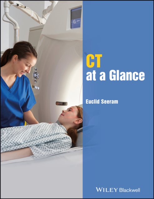
CT at a Glance gets readers quickly up to speed with the core knowledge and competencies required for computed tomography (CT) scanning, as established by the major radiography organizations around the world, including the ASRT and the CAMRT.
CT at a Glance gets readers quickly up to speed with the core knowledge and competencies required for computed tomography (CT) scanning, as established by the major radiography organizations around the world, including the ASRT and the CAMRT.
The principle of tomography is to explore the structure and composition of objects non-destructively along spatial and temporal dimensions, using penetrating radiation, such as X- and gamma-rays, or waves, such as electromagnetic and acoustic waves.
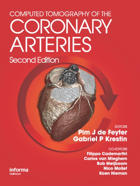
Updated to reflect the notable advances in cardiac computed tomography (CT) imaging, the Second Edition of the best-selling Computed Tomography of the Coronary Arteries provides cardiologists and radiologists with a practical text that explains the basic principles and applications of CT.
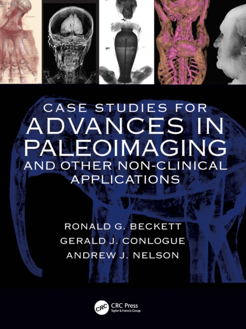
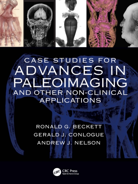
The case studies provided in Case Studies for Advances in Paleoimaging will provide the reader with real-world scenarios and case examples that will help prepare researchers to discover new ways to apply the various modalities associated with the technology.
The case studies provided in Case Studies for Advances in Paleoimaging will provide the reader with real-world scenarios and case examples that will help prepare researchers to discover new ways to apply the various modalities associated with the technology.
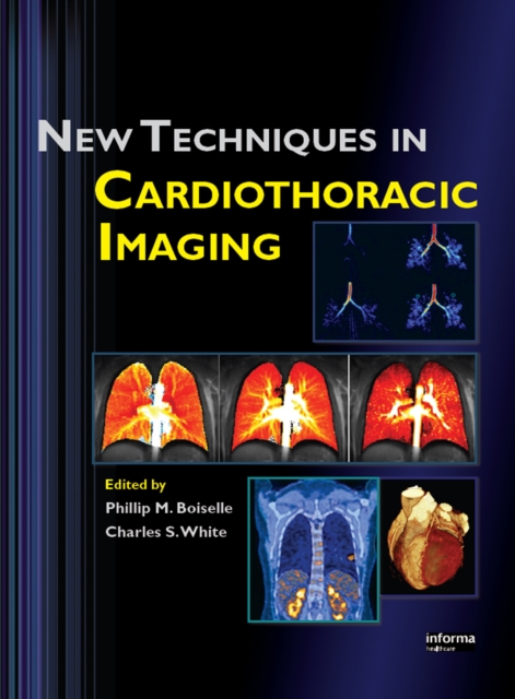
New Techniques in Cardiothoracic Imaging emphasizes emerging methods in computed tomography, magnetic resonance imaging, positron-emission tomography, and similar technology.
Charred, badly decomposed, or mummified corpses, as well as those restrictions forced upon coroners by certain religious sects, often make autopsies impossible to perform.
"e;This book will be useful for all physicians involved in cardiac imaging, whether they are in radiology, nuclear medicine, or cardiology, and should be mandatory for physicians engaged in gated cardiac SPECT.
The principle of tomography is to explore the structure and composition of objects non-destructively along spatial and temporal dimensions, using penetrating radiation, such as X- and gamma-rays, or waves, such as electromagnetic and acoustic waves.
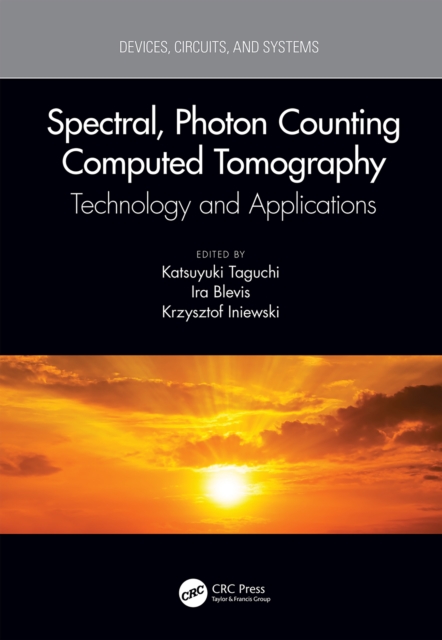
Spectral, Photon Counting Computed Tomography is a comprehensive cover of the latest developments in the most prevalent imaging modality (x-ray computed tomography (CT)) in its latest incarnation: Spectral, Dual-Energy, and Photon Counting CT.
Spectral, Photon Counting Computed Tomography is a comprehensive cover of the latest developments in the most prevalent imaging modality (x-ray computed tomography (CT)) in its latest incarnation: Spectral, Dual-Energy, and Photon Counting CT.
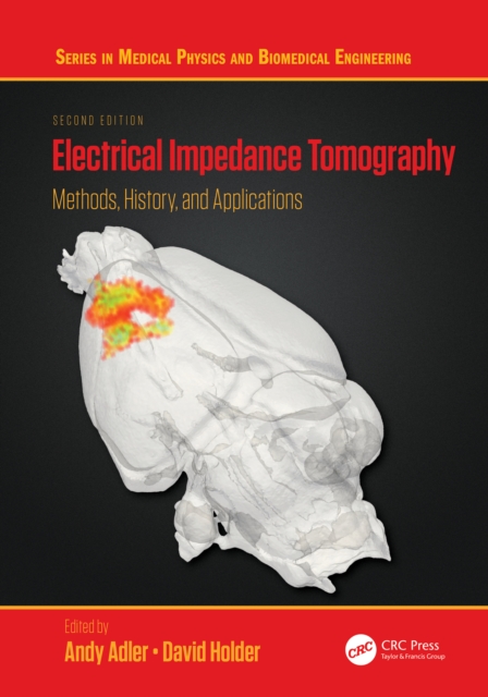
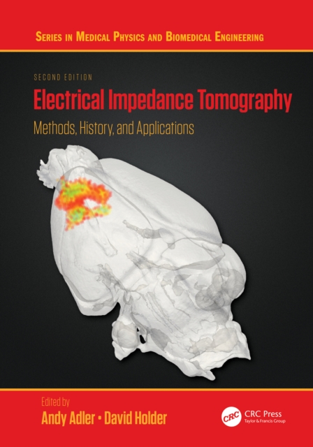
With contributions from leading international researchers, this second edition of Electrical Impedance Tomography: Methods, History and Applications has been fully updated throughout and contains new developments in the field, including sections on image interpretation and image reconstruction.
With contributions from leading international researchers, this second edition of Electrical Impedance Tomography: Methods, History and Applications has been fully updated throughout and contains new developments in the field, including sections on image interpretation and image reconstruction.
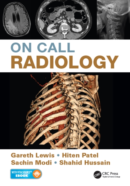
On-Call Radiology presents case discussions on the most common and important clinical emergencies and their corresponding imaging findings encountered on-call.
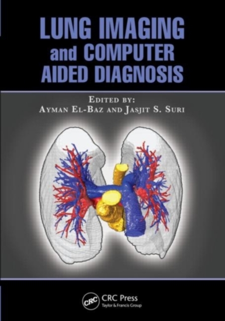
Developing an effective computer-aided diagnosis (CAD) system for lung cancer is of great clinical importance and can significantly increase the patient's chance for survival.
Developing an effective computer-aided diagnosis (CAD) system for lung cancer is of great clinical importance and can significantly increase the patient's chance for survival.