This book systematically covers diagnostic imaging of lung cancers, including epithelial tumors, mesenchymal tumors, and hematolymphoid tumors, with emphasis on relationship between classification, molecular pathology, clinical findings, imaging manifestations and pathological discussion.
This book systematically covers diagnostic imaging of lung cancers, including epithelial tumors, mesenchymal tumors, and hematolymphoid tumors, with emphasis on relationship between classification, molecular pathology, clinical findings, imaging manifestations and pathological discussion.
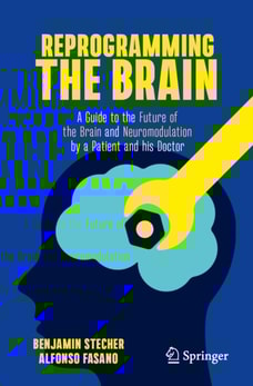
In June 2021, Doctor and Patient decided that time had come to surgically implant two six-inch-long metal alloy spikes all the way through Ben's brain.
This case-based atlas encompasses all aspects of imaging in congenital cardiac defects, cardiac masses, inflammatory and acquired heart diseases, cardiomyopathies and coronary-related pathologies.
This case-based atlas encompasses all aspects of imaging in congenital cardiac defects, cardiac masses, inflammatory and acquired heart diseases, cardiomyopathies and coronary-related pathologies.
This textbook describes the study of radiation, covering the basic concepts and their advanced applications, and highlights the handling of radioisotopes and radiation measurements using various instruments.
This textbook describes the study of radiation, covering the basic concepts and their advanced applications, and highlights the handling of radioisotopes and radiation measurements using various instruments.
The book provides competent assistance to all those affected by Multiple Sclerosis to better understand the disease and educates them about current diagnostic methods and treatment approaches.
This book systematically covers MRI imaging findings of a spectrum of gynaecological diseases, including ovarian cysts, uterine fibroids, cervical cancer, and endometrial cancer, etc.
This comprehensive book features high-quality images, videos, and step-by-step instructions for capturing detailed views of the upper and lower gastrointestinal tract from the oral cavity to the anus.
The book addresses comprehensively the normal and pathological MRI appearance of the structures of the anterior compartment of the knee, potential sources of pain, in a systematic way, on anatomical layers, from superficial to deep, respectively, from prepatellar soft tissues to intra-articular structures (the synovial lining, patellar and trochlear cartilage).
The book addresses comprehensively the normal and pathological MRI appearance of the structures of the anterior compartment of the knee, potential sources of pain, in a systematic way, on anatomical layers, from superficial to deep, respectively, from prepatellar soft tissues to intra-articular structures (the synovial lining, patellar and trochlear cartilage).
The second edition of the well-received book updates the knowledge of clinical treatment, radiobiology, physics, and instrumentations to provide a comprehensive description of stereotactic body radiation therapy (SBRT).
The second edition of the well-received book updates the knowledge of clinical treatment, radiobiology, physics, and instrumentations to provide a comprehensive description of stereotactic body radiation therapy (SBRT).
This book summarizes the recent advancements for visualized medicine in terms of fundamental principles, rapidly emerging techniques and developing frontiers.
This book summarizes the recent advancements for visualized medicine in terms of fundamental principles, rapidly emerging techniques and developing frontiers.
In focussing on the movement and the physiology of the pyloric region, this book uses the methods of anatomy, radiology, gastroscopy, manometry, myoelectric activity and ultrasonography of the pyloric region.