Bioluminescent Imaging: Methods and Protocols distills a wide range of techniques that use bioluminescence imaging as a tool for visualizing and tracking various biological processes.
As fMRI technology has provided invaluable insights into the mechanisms through which the human brain works in healthy individuals and in patients with different neurological and psychiatric conditions, the study of brain function and even the monitoring of the effects of treatment have become more effective and efficient.
With its ability to explore the surface of the sample by means of a local scanning probe and its use of dedicated software allows to be visualize results, atomic force microscopy (AFM) has revolutionized the study of the smallest aspects of life.
Leading experts in the use of MRI explain its basic principles and demonstrate its power to understand biological processes with numerous cutting-edge applications.
As imaging technologies and approaches have evolved, the scope of certain imaging techniques has moved far beyond the production of purely illustrative images or appealing time-lapse movies to providing the scientist with a rich range of ways to measure and quantify the biological process and outcome of gene expression.
In recent years, molecular imaging techniques have grown to be invaluable tools for molecular biology research and, to a more modest extent, clinical medicine.
Now a routine tool in biomedical and life science research, live cell imaging has made major progress enabling this core biochemical, cell, and molecular biology technique to become even more powerful, versatile, and affordable.
Quantitative elucidation of structural, energetic and dynamic aspects of macromolecular interactions is indispensable for understanding the functional activities of biomolecules and their interactions.
Reflecting the expanding field's need for reliable protocols, Fluorescence Spectroscopy and Microscopy: Methods and Protocols offers techniques from a worldwide team of experts on this versatile and vital subject.
This third edition of Electron Microscopy: Methods and Protocols expands upon the previous editions with current, detailed protocols on biological and molecular research techniques based on TEM and SEM as well as other closely related imaging and analytical methods.
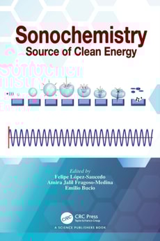
This book explores the most pertinent aspects and advancements in sonochemistry, dedicating nine chapters to fundamentals, synthesis methods, and applications.
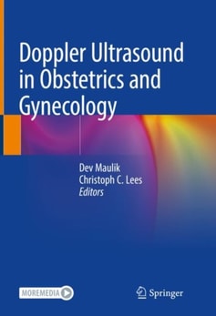
This book, now in a comprehensively revised and expanded third edition, covers the full spectrum of applications of Doppler ultrasound within Obstetrics and Gynecology.
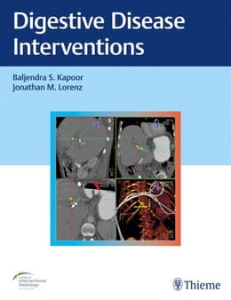
Comprehensively covers the rich spectrum of radiologic digestive disease interventionsGreater understanding of gastrointestinal disease has resulted in an evolving array of minimally invasive and noninvasive techniques.
This book is focused on trigeminal neuralgias and their management, a field in which, during the two past decades, there were numerous advances in comprehension of mechanisms of the disease and diagnosis with imaging, as well as in medical therapies and in surgical treatment.
This book provides an in-depth review of the knowledge of craniospinal arachnoid cysts, with emphasis on epidemiology, genetics, neuroimaging, clinical presentations, and operative management.
This book embarks on a journey never taken before, approaching the imaging of the disease of achalasia with new pathophysiological assumptions in mind, coming from the Chicago Classification of Manometric diagnosis.
This book embarks on a journey never taken before, approaching the imaging of the disease of achalasia with new pathophysiological assumptions in mind, coming from the Chicago Classification of Manometric diagnosis.
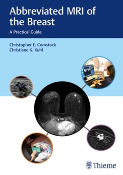
State-of-the-art resource details effective breast MRI techniques for improved screening and diagnosisMagnetic resonance imaging (MRI) of the breast has evolved into an important breast cancer screening tool and major advance in women's health.
Although mammography is the primary method used for breast cancer screening, screening mammography is limited especially in women with dense breasts, which includes nearly 50% of all women in the United States.
Imaging for Students delivers step-by-step guidance to the range of imaging techniques available, providing a clear explanation of how each imaging modality actually works, and including information on the associated risks and hazards.
This book examines deep learning-based approaches in the field of cancer diagnostics, as well as pre-processing techniques, which are essential to cancer diagnostics.
This book examines deep learning-based approaches in the field of cancer diagnostics, as well as pre-processing techniques, which are essential to cancer diagnostics.
This book provides the most up-to-date and comprehensive source of information on all aspects of EEG-fMRI, a neuroimaging technique for synchronous acquisition of electroencephalography (EEG) and functional magnetic resonance imaging (fMRI) data.
This book provides a thorough overview of recent methods using higher level information (object or scene level) for advanced tasks such as image understanding along with their applications to medical images.
Dieses Fachbuch führt in die Besonderheiten des Infrarot und seiner abbildenden Systeme ein und beschreibt die technischen und physikalischen Grundlagen.