The principle of tomography is to explore the structure and composition of objects non-destructively along spatial and temporal dimensions, using penetrating radiation, such as X- and gamma-rays, or waves, such as electromagnetic and acoustic waves.
Highly Commended at the British Medical Association Book Awards 2016Abdominal X-rays for Medical Students is a comprehensive resource offering guidance on reading, presenting and interpreting abdominal radiographs.
Highly Commended at the British Medical Association Book Awards 2016Abdominal X-rays for Medical Students is a comprehensive resource offering guidance on reading, presenting and interpreting abdominal radiographs.
The use of neurovascular ultrasound is of increasing importance in neurological practice, both for radiologists and increasingly by neurologists themselves.
The use of neurovascular ultrasound is of increasing importance in neurological practice, both for radiologists and increasingly by neurologists themselves.
Working practices for Head and Neck (HN) diagnostic and clinical teams have changed dramatically over the past 15 years with highlighted importance on specialist Multidisciplinary Teams (MDT) including radiologists and cytopathologists.
Working practices for Head and Neck (HN) diagnostic and clinical teams have changed dramatically over the past 15 years with highlighted importance on specialist Multidisciplinary Teams (MDT) including radiologists and cytopathologists.
Offers a well-designed approach to imaging musculoskeletal trauma Medical imaging plays an important role in identifying fractures and helping the patient return to regular activities as soon as possible.
Offers a well-designed approach to imaging musculoskeletal trauma Medical imaging plays an important role in identifying fractures and helping the patient return to regular activities as soon as possible.
** New revised second edition now available, with errors corrected and content fully updated ** The second edition of the classic text has been revised and extended to meet the needs of today s practising and training MRI technologists who intend to sit for the American Registry of Magnetic Resonance Imaging Technologists (ARMRIT) examination.
** New revised second edition now available, with errors corrected and content fully updated ** The second edition of the classic text has been revised and extended to meet the needs of today s practising and training MRI technologists who intend to sit for the American Registry of Magnetic Resonance Imaging Technologists (ARMRIT) examination.
Diagnostic Imaging will help medical students, junior doctors, residents and trainee radiologists understand the principles behind interpreting all forms of imaging.
Diagnostic Imaging will help medical students, junior doctors, residents and trainee radiologists understand the principles behind interpreting all forms of imaging.
Rapid acquisition and interpretation of radiographs, portable ultrasound (US) and computed tomography (CT) are now the mainstay of initial successful management of sick and traumatized patients presenting to Accident and Emergency Departments.
Rapid acquisition and interpretation of radiographs, portable ultrasound (US) and computed tomography (CT) are now the mainstay of initial successful management of sick and traumatized patients presenting to Accident and Emergency Departments.
The first complete reference dedicated to the full spectrum of women's imaging topics "e;Women s imaging"e; refers to the use of imaging modalities (X-ray, ultrasound, CT scan, and MRI) available for aiding in the diagnosis and care of such female-centric diseases as cancer of the breast, uterus, and ovaries.
The first complete reference dedicated to the full spectrum of women's imaging topics "e;Women s imaging"e; refers to the use of imaging modalities (X-ray, ultrasound, CT scan, and MRI) available for aiding in the diagnosis and care of such female-centric diseases as cancer of the breast, uterus, and ovaries.
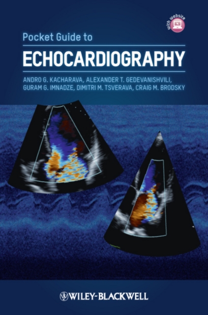
With its easy accessibility, low cost, and ability to deliver, essential bedside information about the cardiac structure and function, echocardiography has become one of the most relied-upon diagnostic tools in clinical medicine.
With its easy accessibility, low cost, and ability to deliver, essential bedside information about the cardiac structure and function, echocardiography has become one of the most relied-upon diagnostic tools in clinical medicine.
Thoroughly and systematically presents the state-of-the-art in the diagnostic uses of radiologic imaging and nuclear medicine in the diagnosis and management of infectious and inflammatory diseases Although our understanding of microorganisms has advanced significantly and antimicrobial therapy has become increasingly available, infection remains a major cause of patient morbidity and mortality.
Thoroughly and systematically presents the state-of-the-art in the diagnostic uses of radiologic imaging and nuclear medicine in the diagnosis and management of infectious and inflammatory diseases Although our understanding of microorganisms has advanced significantly and antimicrobial therapy has become increasingly available, infection remains a major cause of patient morbidity and mortality.
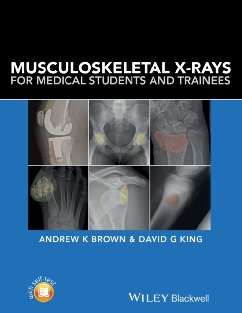
Musculoskeletal X-rays for Medical Students provides the key principles and skills needed for the assessment of normal and abnormal musculoskeletal radiographs.
Musculoskeletal X-rays for Medical Students provides the key principles and skills needed for the assessment of normal and abnormal musculoskeletal radiographs.
Translated into seven languages, Cotton and Williams' Practical Gastrointestinal Endoscopy has for the last 25 years been the basic primer for endoscopy around the world, providing clear, clinical and practical guidance on the fundamentals of endoscopy practice, from patient positioning and safety, how to perform different endoscopic procedures, and the latest in therapeutic techniques and advances in technology.
Translated into seven languages, Cotton and Williams' Practical Gastrointestinal Endoscopy has for the last 25 years been the basic primer for endoscopy around the world, providing clear, clinical and practical guidance on the fundamentals of endoscopy practice, from patient positioning and safety, how to perform different endoscopic procedures, and the latest in therapeutic techniques and advances in technology.
Written by one of the world's most respected cardiologists and designed with the needs of the internist and general clinical cardiologist in mind, this new volume provides clear, accessible guidance on the use of electrocardiography to diagnose and manage cardiovascular disease.
Written by one of the world's most respected cardiologists and designed with the needs of the internist and general clinical cardiologist in mind, this new volume provides clear, accessible guidance on the use of electrocardiography to diagnose and manage cardiovascular disease.
This important volume is the first to address the use of neuroimaging in civil and criminal forensic contexts and to include discussion of prior precedents and court decisions.
This book presents and describes imaging technologies that can be used to study chemical processes and structural interactions in dynamic systems, principally in biomedical systems.
This book presents and describes imaging technologies that can be used to study chemical processes and structural interactions in dynamic systems, principally in biomedical systems.
Expert Report Writing Software provides a step-by-step guide to writing clinically sound and rich psychological reports The Psychological Report Writing Assistant software is a highly interactive program that guides the report writer through all phases of writing a report that is comprehensive, includes integrated interpretation, uses everyday language, and answers the referral questions.
Expert Report Writing Software provides a step-by-step guide to writing clinically sound and rich psychological reports The Psychological Report Writing Assistant software is a highly interactive program that guides the report writer through all phases of writing a report that is comprehensive, includes integrated interpretation, uses everyday language, and answers the referral questions.