More than 4,800 illustrations address common head and neck imaging issues most often faced by radiologists in clinical practiceHead and Neck Imaging Variants delivers more than 375 cases and 4,800 illustrations to help you determine whether a finding is truly abnormal or merely a variant and to avoid common imaging pitfalls.
The third edition of Fundamentals of Mammography assists clinicians to deliver a consistently high-quality service while acquiring the skills needed to provide care at what can be an emotionally difficult time for many patients.
These proceedings cover the talks delivered by experts and pioneers on various aspects of this field: theory; principles of the techniques; instrumentation; data analysis; data interpretation and state-of-the-art studies.
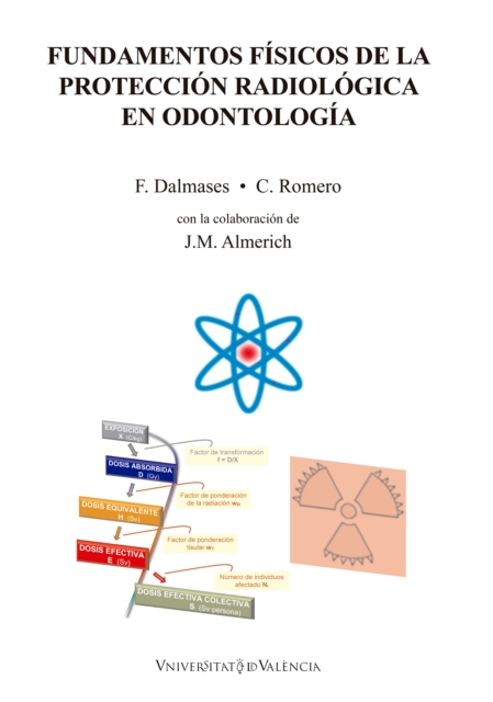
Los rayos X constituyen una herramienta diagnóstica imprescindible en la práctica odontológica actual, a pesar de que comporta una serie de riesgos tanto para el paciente como para el profesional y su entorno.
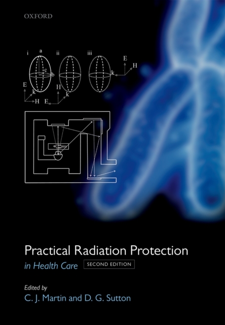
A highly practical reference for health physicists and other professionals, addressing practical problems in radiation protection, this new edition has been completely revised, updated and supplemented by such new sections as log-normal distribution and digital radiography, as well as new chapters on internal radiation dose and the environmental transport of radionuclides.
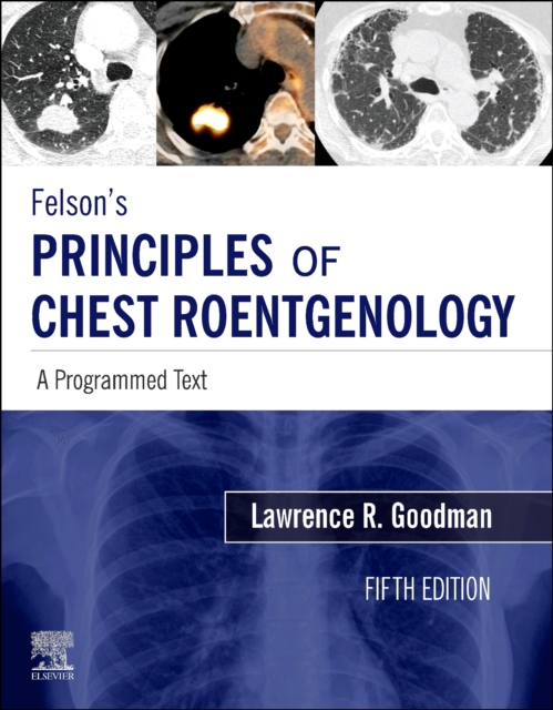
Easy to read, engaging, and highly interactive, Felson's Principles of Chest Roentgenology: A Programmed Text, 5th Edition, has long been the go-to learning resource for medical students, residents, radiologists, and others who order and interpret chest x-rays.
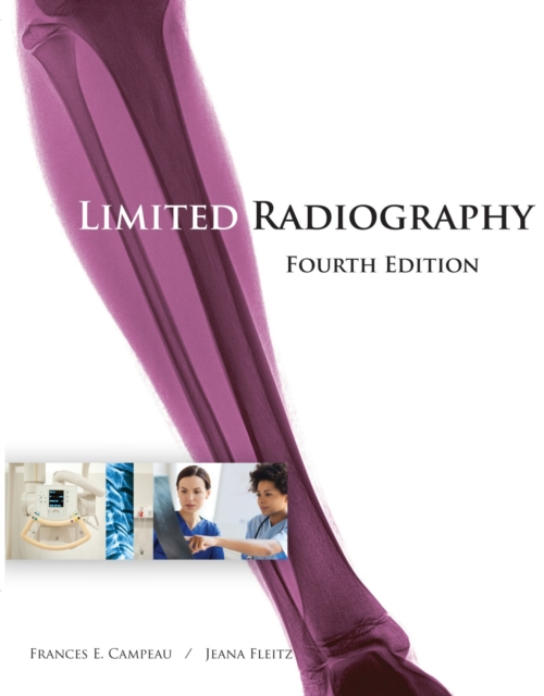
Featuring a new full-color design and packed with illustrations, LIMITED RADIOGRAPHY, 4e is an ideal resource for beginning radiography students and limited radiographer training.
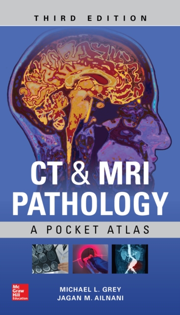
The acclaimed pocket atlas of the most common pathologic conditions seen on CT and MRI - more essential than ever, with new images and cases Designed for quick look-up at the point of care, this concise handbook provides technologists and students with CT and MRI findings of 200 pathologic conditions most often seen in day-to-day practice, along with pertinent clinical information.
Since its first edition in 1980, Essential Physics for Radiographers has earned an international reputation as a clear and straightforward introduction to the physics of radiography.
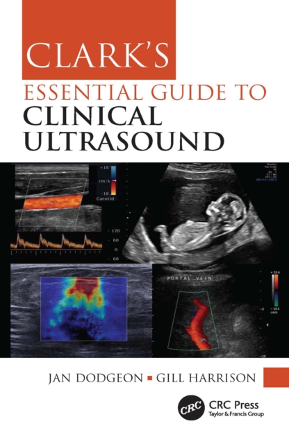
This easy-to-understand pocketbook in the highly respected Clark's stable of diagnostic imaging texts is an invaluable tool for students, sonographers and other ultrasound practitioners, providing practical guidance on clinical ultrasound procedures, summarising current relevant literature and professional body guidelines.
This easy-to-understand pocketbook in the highly respected Clark's stable of diagnostic imaging texts is an invaluable tool for students, sonographers and other ultrasound practitioners, providing practical guidance on clinical ultrasound procedures, summarising current relevant literature and professional body guidelines.
The third edition of Fundamentals of Mammography assists clinicians to deliver a consistently high-quality service while acquiring the skills needed to provide care at what can be an emotionally difficult time for many patients.
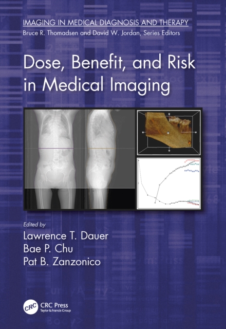
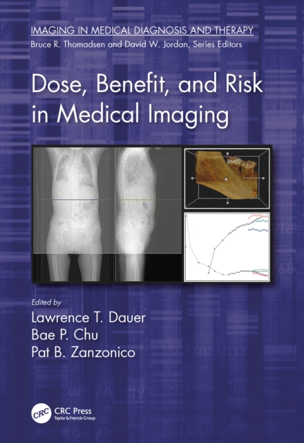
This timely overview of dose, benefit, and risk in medical imaging explains to readers how to apply this information for informed decision-making that improves patient outcomes.
This timely overview of dose, benefit, and risk in medical imaging explains to readers how to apply this information for informed decision-making that improves patient outcomes.
Easy to read, engaging, and highly interactive, Felson's Principles of Chest Roentgenology: A Programmed Text, 5th Edition, has long been the go-to learning resource for medical students, residents, radiologists, and others who order and interpret chest x-rays.
More than 4,800 illustrations address common head and neck imaging issues most often faced by radiologists in clinical practiceHead and Neck Imaging Variants delivers more than 375 cases and 4,800 illustrations to help you determine whether a finding is truly abnormal or merely a variant and to avoid common imaging pitfalls.
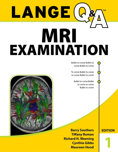
The most complete review available for the ARRT(R) MRI certification examinationLANGE Q&A MRI Examination is a comprehensive review for radiologic technologists preparing to take the ARRT specialty certification exam in magnetic resonance imaging (MRI).
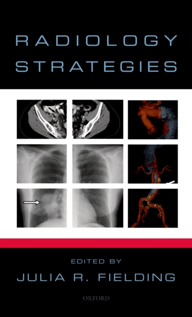
As physicians become more dependent upon radiologic tests for information, it is imperative that they have correct information on how and when to order them.
The Oxford American Handbook of Radiology is a concise, image-rich guide to radiology for non-radiologists who wish to improve their understanding and utilization of imaging as well as their interpretative skills.
Imaging is a critical component in the delivery of radiotherapy to patients with malignancy, and this book teaches the principles and practice of imaging specific to radiotherapy.
Imaging is a critical component in the delivery of radiotherapy to patients with malignancy, and this book teaches the principles and practice of imaging specific to radiotherapy.
This new volume in the Radiotherapy in Practice series provides a comprehensive and evidence-based guide to radiotherapy in the management of children and young people with cancer.
This new volume in the Radiotherapy in Practice series provides a comprehensive and evidence-based guide to radiotherapy in the management of children and young people with cancer.
Challenging Concepts in Interventional Radiology is a cased based guide to the most challenging aspects of interventional radiology and endovascular procedures.
Challenging Concepts in Interventional Radiology is a cased based guide to the most challenging aspects of interventional radiology and endovascular procedures.
Radioisotope therapy is an internal form of radiation used to treat cancer; it may be administered orally or intravenously and represents the nearest treatment option to the 'magic bullet', specifically targeting sites of disease whilst sparing surrounding normal tissues.
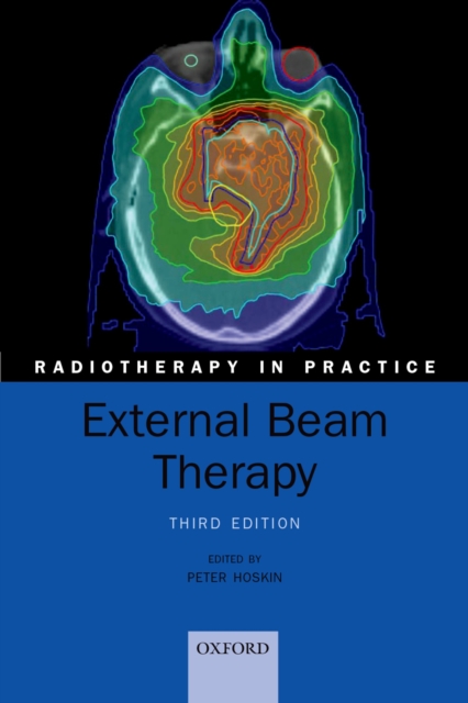
External beam therapy is the most common form of radiotherapy, delivering ionizing radiation such as high-energy x-rays, gamma rays, or electron beams directly into the location of the patient's tumour.
External beam therapy is the most common form of radiotherapy, delivering ionizing radiation such as high-energy x-rays, gamma rays, or electron beams directly into the location of the patient's tumour.
The study of the biology of tumours has grown to become markedly interdisciplinary, involving chemists, statisticians, epidemiologists, mathematicians, bioinformaticians, and computer scientists alongside biologists, geneticists, and clinicians.
The study of the biology of tumours has grown to become markedly interdisciplinary, involving chemists, statisticians, epidemiologists, mathematicians, bioinformaticians, and computer scientists alongside biologists, geneticists, and clinicians.