From the discovery of x-rays in 1895 through the emergence of computed tomography (CT) in the 1970s and magnetic resonance imaging (MRI) in the 1980s, non-invasive imaging has revolutionized the practice of medicine.
The Radiological Sciences Dictionary is a rapid reference guide for all hospital staff employed in diagnostic imaging, providing definitions of over 3000 keywords as applied to the technology of diagnostic radiology.
Providing the most comprehensive, up-to-date coverage of this exciting biomedical field, Handbook of Photomedicine gathers together a large team of international experts to give you a complete account of the application of light in healthcare and medical science.
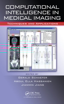
CI Techniques & Algorithms for a Variety of Medical Imaging SituationsDocuments recent advances and stimulates further researchA compilation of the latest trends in the field, Computational Intelligence in Medical Imaging: Techniques and Applications explores how intelligent computing can bring enormous benefit to existing technology in medical
Propelling quantitative MRI techniques from bench to bedside, Quantitative MRI in Cancer presents a range of quantitative MRI methods for assessing tumor biology.
To improve efficiency and reduce administrative costs, healthcare providers, insurance companies, and governments are increasingly using integrated electronic health record (EHR) and picture archiving and communication systems (PACS) to manage patients' medical information.
Improve the Accurate Detection and Diagnosis of Cancer and Other DiseasesDespite the expansion of the CAD field in recent decades, there is currently no single book dedicated to the development and use of CAD systems.
Radiation Protection in Medical Imaging and Radiation Oncology focuses on the professional, operational, and regulatory aspects of radiation protection.
While there are many excellent texts focused on clinical medical imaging, there are few books that approach in vivo imaging technologies from the perspective of a scientist or physician-scientist using, or interested in using, these techniques in research.
Stem Cell Labeling for Delivery and Tracking Using Noninvasive Imaging provides a comprehensive overview of cell therapy imaging, ranging from the basic biology of cell therapeutic choices to the preclinical and clinical applications of cell therapy.
Targeted Molecular Imaging covers the development of novel diagnostic approaches that use an imaging probe and agent to noninvasively visualize cellular processes in normal and disease states.
From the Watching of Shadows: The Origins of Radiological Tomography presents the first complete history of body imaging by discrete sections, from its earliest beginnings around 1920 to modern times.
Physiological optical imaging is a group of emerging technologies that aim to provide healthcare practitioners and biomedical researchers with information about tissue physiology or pathophysiology using approaches different from traditional medical imaging (PET, ultrasound, MRI, X-ray, or CT scan).
Computational biomechanics is an emerging research field that seeks to understand the complex biomechanical behaviors of normal and pathological human joints to come up with new methods of orthopedic treatment and rehabilitation.
This book examines Dynamic Light Scattering (DLS) and its derivatives Laser Doppler Flowmetry (LDF), Diffusing Wave Spectroscopy (DWS), Laser Speckle Contrast Imaging (LSCI), and Doppler Optical Coherence Tomography (OCT) for characterizing particle motion in turbid mediums like suspensions and solutions.
Linear Accelerators for Radiation Therapy, Second Edition focuses on the fundamentals of accelerator systems, explaining the underlying physics and the different features of these systems.
Through a biophysical approach, Electromagnetic Fields in Biology and Medicine provides state-of-the-art knowledge on both the biological and therapeutic effects of Electromagnetic Fields (EMFs).
This book examines Dynamic Light Scattering (DLS) and its derivatives Laser Doppler Flowmetry (LDF), Diffusing Wave Spectroscopy (DWS), Laser Speckle Contrast Imaging (LSCI), and Doppler Optical Coherence Tomography (OCT) for characterizing particle motion in turbid mediums like suspensions and solutions.
With every chapter revised and updated, Physics for Diagnostic Radiology, Third Edition continues to emphasise the importance of physics education as a critical component of radiology training.
Due to the increasing number of digital mammograms and the advent of new kinds of three-dimensional x-ray and other forms of medical imaging, mammography is undergoing a dramatic change.
The unprecedented potential of nanotechnology for early detection, diagnosis, and personalized treatment of diseases has found application in every biomedical imaging modality.
Combining facets of health physics with medicine, An Introduction to Radiation Protection in Medicine covers the background of the subject and the medical situations where radiation is the tool to diagnose or treat human disease.
Modern medical imaging and radiation therapy technologies are so complex and computer driven that it is difficult for physicians and technologists to know exactly what is happening at the point-of-care.
The sixth edition of this internationally successful text includes the many positive advances in radiation oncology that have occurred over the past decade, and which continue to keep radiation at the cutting edge of cancer therapy.
Conventional computed tomography (CT) techniques employ a narrow array of x-ray detectors and a fan-shaped x-ray beam to rotate around the patient to produce images of thin sections of the patient.
Understand Quantitative Radiobiology from a Radiation Biophysics PerspectiveIn the field of radiobiology, the linear-quadratic (LQ) equation has become the standard for defining radiation-induced cell killing.
The sixth edition of this internationally successful text includes the many positive advances in radiation oncology that have occurred over the past decade, and which continue to keep radiation at the cutting edge of cancer therapy.
Rapid advances in nanotechnology have enabled the fabrication of nanoparticles from various materials with different shapes, sizes, and properties, and efforts are ongoing to exploit these materials for practical clinical applications.
Fundamentals of MRI: An Interactive Learning Approach explores the physical principles that underpin the technique of magnetic resonance imaging (MRI).
Spectroscopic Tools and Techniques for Analysis of Dental Materials: Current Trends introduces the dental materials and spectroscopic techniques applied for the analysis of such materials, including ceramic, metallic, polymeric and composites.
This concise yet comprehensive primer on ionising radiation protection includes chapters on foundational nuclear physics, radiation units, the biological effects of radiation exposure, radiation risk and epidemiological studies, internal and external radiation hazards, background radiation, radioactive waste management, and radiation detection.
From the Watching of Shadows: The Origins of Radiological Tomography presents the first complete history of body imaging by discrete sections, from its earliest beginnings around 1920 to modern times.
Covering topics in Radiobiology, Modern Physics, Medical Imaging and Radiation Therapy, Foundations of Medical Physics serves as an introduction to the field of Medical Physics, or Radiation Oncology Physics.
This book provides readers with an accessible and up-to-date introduction to the field of low-field MRI, which is currently seeing a resurgence in both research and commercial activity.
This book focuses on a systematic introduction to the knowledge of mathematics and physics of electroencephalogram (EEG) and discusses an in-depth application of EEG and the development of new methods and technologies for mining and analyzing EEG.