This book covers various facets of nanomaterials and their applications including low-dimensional materials along with discussions on in vitro cell imaging, bioanalyses, UV laser applications of scheelite-type nanomaterials, and nanosized cyanobridged metal-organic frameworks, including high spin transition metal ions.
An essential guide to honing and developing skills in the review of musculoskeletal MRI studiesDesigned specifically for orthopedic surgeons involved in the review of musculoskeletal MRIs, this book enables clinicians to develop a systematic approach to the interpretation of MRI studies.
Learn the fundamental principles and clinical applications of the diagnostic and risk assessment imaging test of choice for prostate cancer: prostate MRIAlthough prostate cancer is the second leading cause of cancer death in men in the USA, it can be treated successfully if detected early.
MRI Essentials for the Spine Specialist is a comprehensive textbook that details the complex MRI anatomy of the spine and the spectrum of pathological findings in patients with spinal disorders.
The definitive guide to MRI for musculoskeletal abnormalitiesDifferential Diagnosis in Musculoskeletal MRI is a unique desk reference offering extensive descriptions of MRI findings that enable radiologists to more easily diagnose a wide range of musculoskeletal conditions.
Gain crucial diagnostic skills with this image-rich guide to clinical MR interpretationEssentials of Clinical MR provides readers with the must-have background they need to read magnetic resonance images and make successful clinical diagnoses.
Organized by findings to reflect how radiologists really work, this abundantly illustrated book offers more than 2,000 magnetic resonance images depicting commonly seen congenital and acquired disorders, as well as many rare and unusual cases.
An introduction to the premier clinical imaging field strength for MRHere is the first textbook to present a practical overview of the basic principles and clinical applications for 3 tesla (3 T) MR imaging.
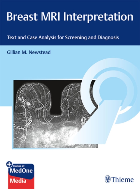
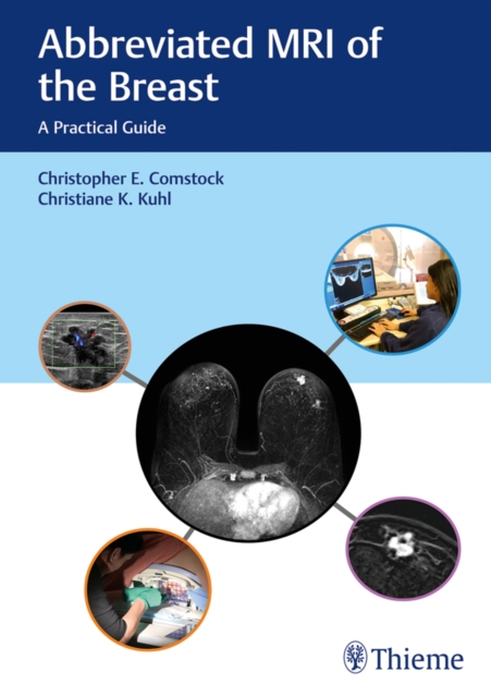
State-of-the-art resource details effective breast MRI techniques for improved screening and diagnosisMagnetic resonance imaging (MRI) of the breast has evolved into an important breast cancer screening tool and major advance in women's health.
Although mammography is the primary method used for breast cancer screening, screening mammography is limited especially in women with dense breasts, which includes nearly 50% of all women in the United States.
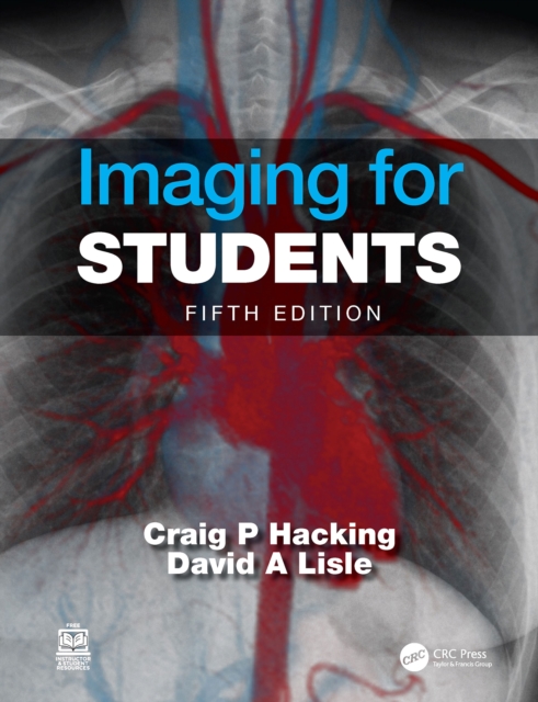
Imaging for Students delivers step-by-step guidance to the range of imaging techniques available, providing a clear explanation of how each imaging modality actually works, and including information on the associated risks and hazards.
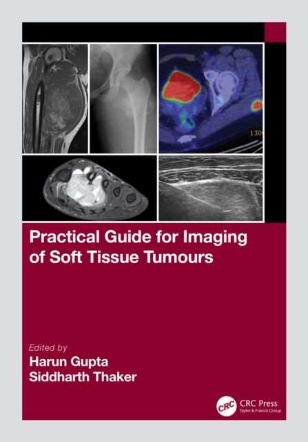
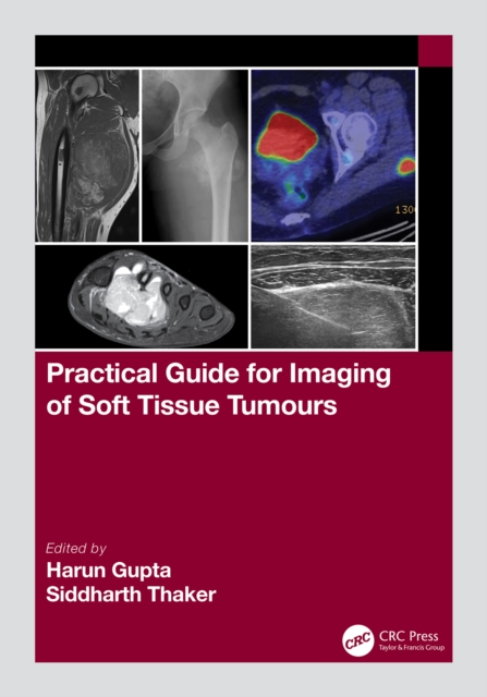
Soft tissue tumours are extremely common although difficult to understand due to a large number of sub-types, leading to a significant increase in their imaging in the last decade.
Soft tissue tumours are extremely common although difficult to understand due to a large number of sub-types, leading to a significant increase in their imaging in the last decade.
Since the development of the first clinical MRI contrast agent, Gd(DTPA) (or Magnevist(R)) in the early 1980s, another three linear and three macrocyclic (eg.
- Provides state-of-the-art coverage of CMR technologies and guidelines, including basic principles, imaging techniques, ischemic heart disease, right ventricular and congenital heart disease, vascular and pericardium conditions, and functional cardiovascular disease.
This is the first book that presents a comprehensive introduction to and overview of electro-magnetic tissue property imaging techniques using MRI, focusing on Magnetic Resonance Electrical Impedance Tomography (MREIT), Electrical Properties Tomography (EPT) and Quantitative Susceptibility Mapping (QSM).
Imaging for Students delivers step-by-step guidance to the range of imaging techniques available, providing a clear explanation of how each imaging modality actually works, and including information on the associated risks and hazards.
This must-have book is the first self-contained summary of recent developments in the field of microscale nuclear magnetic resonance hardware, covering the entire technology from miniaturized detectors, the signal processing chain, and detection sequences.
This must-have book is the first self-contained summary of recent developments in the field of microscale nuclear magnetic resonance hardware, covering the entire technology from miniaturized detectors, the signal processing chain, and detection sequences.
This book is concerned with imaging of the interior of the human body using signals originating from magnetic moments of hydrogen nuclei and detected using a technique known as magnetic resonance.
A host of imaging techniques are available to clinical cardiologists, including nuclear imaging, echocardiography, computerized tomography, and magnetic-resonance imaging.
The combination of two leading imaging techniques - magnetic resonance imaging and positron emission tomography - is poised to have a large impact and has recently been a driver of research and clinical applications.
The combination of two leading imaging techniques - magnetic resonance imaging and positron emission tomography - is poised to have a large impact and has recently been a driver of research and clinical applications.
As a practical reference guide for designing and performing experiments, this book focuses on the five most common classes of contrast agents for MRI namely gadolinium complexes, chemical exchange saturation transfer agents, iron oxide nanoparticles, manganese complexes and fluorine contrast agents.
Spin relaxation parameters, although difficult to interpret, are attracting interest in NMR as these parameters are capable of yielding both structural and dynamic information.
As a practical reference guide for designing and performing experiments, this book focuses on the five most common classes of contrast agents for MRI namely gadolinium complexes, chemical exchange saturation transfer agents, iron oxide nanoparticles, manganese complexes and fluorine contrast agents.
This is the first book that presents a comprehensive introduction to and overview of electro-magnetic tissue property imaging techniques using MRI, focusing on Magnetic Resonance Electrical Impedance Tomography (MREIT), Electrical Properties Tomography (EPT) and Quantitative Susceptibility Mapping (QSM).
Providing the first comprehensive book on the current state of hyperpolarized Xenon-129 NMR and MRI, this book is guaranteed to appeal to a wide range of scientists interested in this growing field.
Spin relaxation parameters, although difficult to interpret, are attracting interest in NMR as these parameters are capable of yielding both structural and dynamic information.
Fundamentals of MRI: An Interactive Learning Approach explores the physical principles that underpin the technique of magnetic resonance imaging (MRI).
The opening chapter of The Essential Guide to Magnetic Resonance presents applications of Fluorine-19 Magnetic Resonance Imaging (19F MRI) in cancer research, tissue metabolomics, quantification of drug delivery, cellular tracking, tissue pH measurements and detection of 19F labeled cells.
The field of magnetic resonance imaging (MRI) has developed rapidly over the past decade, benefiting greatly from the newly developed framework of compressed sensing and its ability to drastically reduce MRI scan times.
Based on research and clinical trials, this book details the latest research in magnetic resonance imaging (MRI) tagging technology related to heart mechanics.
MRI techniques have been recently introduced for non-invasive qualification of regional myocardial mechanics, which is not achievable with other imaging modalities.