Maintaining the first edition's unique parallel to the strategy used by pathologists and pulmonologists to arrive at a patient's diagnosis in daily practice, Diagnostic Pulmonary Pathology starts with the patient and their biopsy findings, directing the pathologist or clinician to the proper diagnosis.
An ideal text for radiation oncologists, hematologist-oncologists, and radiologists, Image-Guided Radiotherapy and Functional Imaging in Modern Lymphoma Management is the foremost source for information on the increasingly important subject of image guided radiation therapy (IGRT) and its crucial role in the clinical evolution of high-precision ion
The Ischemic Penumbra presents the current status of concepts and research on this topic and identifies the latest methods for clinicians to quickly and efficiently recognize viable cerebral tissue for enhanced stroke management.
With the emergence of genetically manipulated laboratory mice as one of the most powerful tools for neuroscientists, imaging techniques capable of providing anatomical and functional information of small animals have become extremely important.
Kinematic MRI refers to imaging a joint through a range of motion to examine the interactions between the soft tissue and osseous anatomy that comprise the joint.
Image registration is the process of systematically placing separate images in a common frame of reference so that the information they contain can be optimally integrated or compared.
Magnetic Resonance Procedures: Health Effects and Safety is the first authoritative text on MR procedures and its associated health and safety concerns written by noted radiologists, physicists, and scientists with expertise in the field.
Vulnerable plaque development is the result of a complex series of molecular and cellular events involving inflammation, apoptosis, rupture, and thrombosis.
Providing a clear foundation as to what a clot is, how it forms, and the most recent approaches to treatment, Thrombus and Stroke is an all-inclusive resource covering: The fundamental science of clot formation and the pharmacokinetics of thrombolysis The clinical impact of thrombus as it pertains to stroke and the most recent clinical and minimally-invasive approaches to this disorder Current pharmacological interventions and the latest findings on neurovascular disease Clinical management of stroke Thrombogenicity of vascular implants In vivo stroke modeling Methods to assess the efficacy of anticoagulation therapy From the latest imaging techniques to endovascular treatments, this comprehensive, advanced volume provides critical data needed for diagnosis and treatment.
Musculoskeletal Radiology is a single-source guide encompassing all of musculoskeletal imaging, examining classical diseases, as well as modern interpretations of disease.
New Techniques in Cardiothoracic Imaging emphasizes emerging methods in computed tomography, magnetic resonance imaging, positron-emission tomography, and similar technology.
MRI-Guided Focused Ultrasound Surgery will be the first publication on this new technology, and will present a variety of current and future clinical applications in tumor ablation treatment.
The only source to fully cover every aspect of brain embolism, this guide analyzes the causes, symptoms, diagnosis, and management of this disorder-providing a detailed overview of major topics pertinent to embolism including donor sources, recipient sites, embolic material, recipient brain supply arteries, vascular and brain pathology, and the tre
Offering lower toxicity and higher accuracy than conventional therapies, this source offers illustrative coverage of this new method to treat tumors associated with brain, breast, lung, and neuroendocrine cancers.
This comprehensive reference discusses the use of ultrasound, computed tomography (CT), magnetic resonance imaging (MRI), and radioisotopes in the imaging of the adult and pediatric urinary tract.
With 2300 radiological images dispersed throughout the text, this source provides an expansive armamentarium of case studies and examples showcasing both common and uncommon liver pathologies.
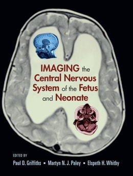
This reference provides an authoritative overview of the role of ultrasonography and MR imaging technologies in the examination and assessment of the central nervous system of the fetus and neonate.
Exploring a technology that is significantly impacting the noninvasive evaluation of the physiology and anatomy of tumors, as well as the diagnosis of infectious processes and cardiac diseases, this source presents recent advances and clinical applications of sequential, single session single-photon emission computed tomography and computed tomography imaging.
This reference documents state-of-the-art trends and advancements in the utilization imaging modalities for the analysis of bones and their surrounding soft tissues, including muscles, tendons, ligaments, nerves, and blood vessels.
The ability of molecular and cellular imaging to track the survival, migration, and differentiation of cells in vivo as well as monitor particular gene expression in living subjects is rapidly moving from the research laboratory into daily clinical settings.
Although there are numerous books on drug metabolism, Radiotracers in Drug Development is unique in explaining how radiotracers are used to elucidate a drug's absorption, distribution, metabolism, and excretion (ADME).
The treatment of a patient with radiation therapy is planned to find the optimal way to treat a tumour while minimizing the dose received by the surrounding normal tissues.
The treatment of a patient with radiation therapy is planned to find the optimal way to treat a tumour while minimizing the dose received by the surrounding normal tissues.
Appraising cancer as a major medical market in the 2010s, Wall Street investors placed their bets on single-technology treatment facilities costing $100-$300 million each.
Appraising cancer as a major medical market in the 2010s, Wall Street investors placed their bets on single-technology treatment facilities costing $100-$300 million each.
From x-rays to lasers to magnetic resonance imaging, developments in basic physics research have been transformed into medical technologies for imaging, surgery and therapy at an ever-accelerating pace.
From x-rays to lasers to magnetic resonance imaging, developments in basic physics research have been transformed into medical technologies for imaging, surgery and therapy at an ever-accelerating pace.
The Manual of Venous and Lymphatic Diseases constitutes a concise but comprehensive and contemporary description of the nature and management of venous and lymphatic diseases.
The Manual of Venous and Lymphatic Diseases constitutes a concise but comprehensive and contemporary description of the nature and management of venous and lymphatic diseases.
This book, written by premier authors in the field of OCT intravascular imaging, covers the best practices for using OCT to provide high resolution cross-sectional viewing for atherosclerotic plaque assessment, stent strut coverage and apposition, assessment in stent restenosis evaluation, and PCI guide and optimization.
This book, written by premier authors in the field of OCT intravascular imaging, covers the best practices for using OCT to provide high resolution cross-sectional viewing for atherosclerotic plaque assessment, stent strut coverage and apposition, assessment in stent restenosis evaluation, and PCI guide and optimization.
This book is an informed, educational and abundantly illustrated guide to the imaging knowledge that medical students in the clinical years of their undergraduate studies will be required to get to know, understand and recall in order to negotiate successfully their finals exams.
This book is designed to convey as much information as possible in a concise and simple way to make it suitable for students, researchers and clinical medical physicists.
Tumors and cancers are insidious diseases characterized by uncontrolled growth of abnormal cells that extend beyond their usual boundaries and disrupt the normal functions of affected organs.
Tumors and cancers are insidious diseases characterized by uncontrolled growth of abnormal cells that extend beyond their usual boundaries and disrupt the normal functions of affected organs.
In the past two decades, significant advances in magnetic resonance microscopy (MRM) have been made possible by a combination of higher magnetic fields and more robust data acquisition technologies.
In the past two decades, significant advances in magnetic resonance microscopy (MRM) have been made possible by a combination of higher magnetic fields and more robust data acquisition technologies.
Specifically aimed at candidates taking their higher exams in clinical medicine (such as the Boards Examinations in the United States and Membership of The Royal College of Physicians in the United Kingdom), Cross-sectional Diagnostic Imaging focuses on cross-sectional imaging (computed tomography, magnetic resonance, and ultrasound, and also includes nuclear medicine isotope imaging) since these cross-sectional techniques have become integral to modern clinical practice.
Specifically aimed at candidates taking their higher exams in clinical medicine (such as the Boards Examinations in the United States and Membership of The Royal College of Physicians in the United Kingdom), Cross-sectional Diagnostic Imaging focuses on cross-sectional imaging (computed tomography, magnetic resonance, and ultrasound, and also includes nuclear medicine isotope imaging) since these cross-sectional techniques have become integral to modern clinical practice.
This is a comprehensive, large-format review text with complete answers for the American national examination of the Registry of Diagnostic Medical Sonographers (RDMS).