Dieses Praxisbuch fasst den aktuellen „State of the Art“ der peripheren arteriellen Interventionen in kompakter und übersichtlicher Weise für den Leser zusammen.
The articles in this volume cover the various radiosurgical techniques used to treat benign and malignant intracranial tumors, cavernous malformations, and functional disorders, as well as a wide array of specific details on medical physics, neuroimaging, and anesthetic support.
Al igual que en la edición anterior, la nueva edición incluye un número significativo de vídeos (407) y animaciones intercaladas en el texto (88), que reflejan situaciones reales y que proporcionan al lector una vía de aprendizaje práctico necesaria para la correcta utilización de la ecografía.
Designed to help you quickly learn or review normal anatomy and confirm variants, Imaging Anatomy: Ultrasound, second edition, is the ultimate reference worldwide, keeping you current within the fast-changing field of ultrasound imaging through comprehensive coverage of sonographic anatomy for head and neck, musculoskeletal, abdomen and pelvis, obstetrics and embryology, neonatal head, and vascular.
This monograph, now in its 2nd edition with 31 new chapters and significant updates, is the first book of its kind written specifically for graduate students and clinicians.
This book presents 100 challenging cases encountered in vascular surgery practice that were selected from the author's vascular registry of 7,000 vascular reconstructions (endovascular and open).
This edited book focuses on the application of patient care within the three specialisms: diagnostic radiography (including fluoroscopy, computed tomography, breast imaging, ultrasound, and magnetic resonance imaging), radiotherapy and oncology, and nuclear medicine and molecular imaging.
A practical, case-based guide on how to perform minimally invasive, image-guided procedures for pain managementMinimally-invasive techniques with fewer complications are continually being developed to provide relief to patients with debilitating, unrelenting pain.
Physiological optical imaging is a group of emerging technologies that aim to provide healthcare practitioners and biomedical researchers with information about tissue physiology or pathophysiology using approaches different from traditional medical imaging (PET, ultrasound, MRI, X-ray, or CT scan).
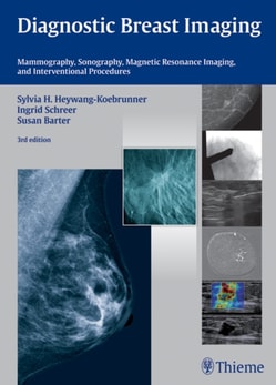
Encompassing the entire spectrum of breast imaging and diagnostics, this acclaimed text provides a systematic and pragmatic guide for all clinicians involved in diagnosing breast disease.
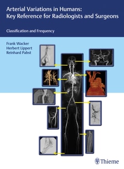
A one-of-a-kind reference and atlas to variations in the human arterial systemBased on the landmark work Arterial Variations in Man: Classification and Frequency by Lippert and Pabst, this atlas presents the full range of arterial variations that occur in the human body.
This book provides a comprehensive overview of the development of the human central nervous system (CNS) in the context of its many developmental disorders due to genetic, environmental and hypoxic/ischaemic causes.
Highly specialized structures, microanatomy of individual components, and overall structural density make the head and neck one of the most challenging areas in radiology.
An image-rich neuroradiology reference and board prep from renowned expertsNeuroradiology: The Essentials with MR and CT, Second Edition, written by world-renowned neuroradiologist and MRI pioneer Val Runge, builds on the acclaimed prior edition.
More than 150 spinal radiology cases deliver the best board review possiblePart of McGraw-Hill Education's Radiology Case Review Series, this unique resource challenges you to look at a group of images, determine the diagnosis, answer related questions, and gauge your knowledge by reviewing the answer.
The Student-to-Student, Step-by-Step Guide to Radiology Clerkship SuccessThis survival guide for the radiology wards teaches you how to read and present normal and abnormal x-rays, CTs, and MRIs for the most commonly seen diseases and disorders.
The book provides detailed information on breast cancer and covers all the aspects of this rapidly spreading disease, such as applied anatomy and physiology, causative factors, various Investigations to reach a concise, definitive and complete diagnosis.
An in-depth guide to upper and lower extremity anatomy based on the latest imaging techniquesWhile the study of anatomy plays a fundamental role in the practice of medicine, most textbooks do not rely on modern imaging and post-processing methods to depict and increase its understanding.
This book offers a comprehensive exploration of brain tumors, beginning with a foundational understanding of their pathophysiology and extending through the latest advancements in diagnosis and treatment.