Vortex Formation in the Cardiovascular System will recapitulate the current knowledge about the vortex formation in the cardiovascular system, from mechanics to cardiology.
This official textbook of the Society of Neurocritical Care (SNCC) comprises a comprehensive review of all possible neurologic conditions that one may encounter in the practice of neuro-intensive care.
Following the recent release of the new Royal College of Radiology Curriculum, the highly successful text, Rapid Review of Radiology has been updated and expanded to ensure full coverage of the new curriculum.
Following the recent release of the new Royal College of Radiology Curriculum, the highly successful text, Rapid Review of Radiology has been updated and expanded to ensure full coverage of the new curriculum.
This book introduces the field of molecular diagnostics, tracing its evolution from the earliest days of genetic research to cutting-edge advancements.
This handbook is a practical guide for interventional cardiology procedures, providing a fast-access reference tool to be consulted in daily practice in the cath lab.
This handbook is a practical guide for interventional cardiology procedures, providing a fast-access reference tool to be consulted in daily practice in the cath lab.
This book introduces the field of molecular diagnostics, tracing its evolution from the earliest days of genetic research to cutting-edge advancements.
This fully updated volume brings together synapse development methods that span from the macroscopic to the nanoscale in order to foster novel perspectives and encourage interdisciplinary integration.
This fully updated volume brings together synapse development methods that span from the macroscopic to the nanoscale in order to foster novel perspectives and encourage interdisciplinary integration.
The second edition of the esteemed atlas provides an in-depth exploration of craniofacial anatomy, enhanced by images of real cadavers that vividly depict the details of human anatomy.
This book delves into the intricate journey of neuroendocrine evolution, from its rudimentary origins in single-celled organisms to the complex systems found in mammals.
The "e;Neuroendocrine and Oral Cancers: An Interdisciplinary Approach"e; is the fourteenth volume of the "e;Interdisciplinary Cancer Research"e; series, publishes comprehensive volume on diagnosis and treatment of neuroendocrine tumors and oral cancers.
The "e;Neuroendocrine and Oral Cancers: An Interdisciplinary Approach"e; is the fourteenth volume of the "e;Interdisciplinary Cancer Research"e; series, publishes comprehensive volume on diagnosis and treatment of neuroendocrine tumors and oral cancers.
The "e;Brain Tumors: An Interdisciplinary Approach"e; is the thirteenth volume of the "e;Interdisciplinary Cancer Research"e; series, publishes comprehensive volume on diagnosis and treatment of brain tumors.
The "e;Brain Tumors: An Interdisciplinary Approach"e; is the thirteenth volume of the "e;Interdisciplinary Cancer Research"e; series, publishes comprehensive volume on diagnosis and treatment of brain tumors.
The "e;Cutaneous Cancers: An Interdisciplinary Approach"e; is the fifteenth volume of the "e;Interdisciplinary Cancer Research"e; series, publishes comprehensive volume on diagnosis and treatment of cutaneous cancers.
The "e;Cutaneous Cancers: An Interdisciplinary Approach"e; is the fifteenth volume of the "e;Interdisciplinary Cancer Research"e; series, publishes comprehensive volume on diagnosis and treatment of cutaneous cancers.
This book describes the role of radioactivity in the living world and highlights the many aspects of the impact of ionizing radiation on biological systems.
Primer on Radiation Oncology Physics: Video Tutorials with Textbook and Problems, now in its second edition, provides over 60 tutorial videos (each 15-20 minutes in length) with a companion text and is the most complete and effective introduction to medical physics available.
This Challenging Cases book provides a structured pathway to help unlock the mysteries of fibrosing Interstitial Lung Diseases (ILDs), guiding experts and beginners alike through the complexities and essential information of fibrosing ILD, interstitial pneumonias (IPs), and HRCT findings and signs.
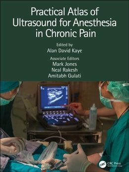
The use of ultrasound to help clinicians specializing in the treatment of chronic pain has expanded greatly over recent years; this illustrated text from a team of experts gives both the experienced practitioner and the trainee a state-of-the-art course in safe and effective techniques.
Updated to reflect the latest scientific advances and technologies in the diagnosis and treatment of pleural diseases, this new Second Edition explores the structure and function of these diseases and malignancies, from tuberculosis and asbestos to pleurisy and pneumothorax.
Formulated by members of the International Scientific Committee of Radionuclides in Nephro-urology (ISCORN), Functional Imaging in Nephro-urology is not a textbook on uronephrology or radionuclides in nephro-urology, or even a book on new techniques in imaging.
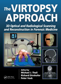
Charred, badly decomposed, or mummified corpses, as well as those restrictions forced upon coroners by certain religious sects, often make autopsies impossible to perform.
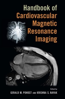
Cardiovascular Magnetic Resonance (CMR) is well established in clinical practice for the diagnosis and management of a wide array of cardiovascular diseases.
Written by internationally known experts in the field, Stereotactic Radiosurgery and Stereotactic Body Radiation Therapy examines one of the fastest-developing subspecialties within radiation oncology.
Comprised of chapters carefully selected from CRC's best-selling engineering handbooks, volumes in the Principles and Applications in Engineering series provide convenient, economical references sharply focused on particular engineering topics and subspecialties.
Image registration is the process of systematically placing separate images in a common frame of reference so that the information they contain can be optimally integrated or compared.
This reference provides an authoritative overview of the role of ultrasonography and MR imaging technologies in the examination and assessment of the central nervous system of the fetus and neonate.
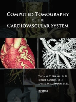
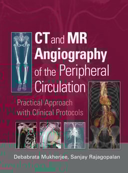
This text discusses the basic aspects of multislice CT angiography with chapters on technical principles, basic scan technique for peripheral vascular imaging with multislice CT, image reconstruction with multislice CT, radiation doses, and contrast agent administration.
Reflecting the increased importance of the collaborations between radiation oncology and informatics professionals, Informatics in Radiation Oncology discusses the benefits of applying informatics principles to the processes within radiotherapy.
Experiments in Nuclear Science is an introductory-level laboratory manual providing hands-on opportunities for developing insights into the origins and properties of nuclear radiations, their interactions with matter, their detection and measurement, and their applications in the physical and life sciences.
Edited by a renowned international expert in the field, Nuclear Medicine Physics offers an up-to-date, state-of-the-art account of the physics behind the theoretical foundation and applications of nuclear medicine.
Radiosensitizers and Radiochemotherapy in the Treatment of Cancer catalogs and describes the mechanism of action for entities characterized as radiosensitizers.
Covering both noninvasive and surgical treatment alternatives, Critical Limb Ischemia defines practical guidelines for a multidisciplinary approach to critical limb ischemia and follows a step-by-step description of the latest techniques.
The field of medical imaging has been revolutionized by new techniques in powerful computations, image processing, and modalities such as Computer-Aided Tomography (CAT) and Magnetic Resonance Imaging (MRI), among others.
Exploring novel methods in endovascular intervention, this reference provides detailed coverage of techniques to avoid, manage, and control complications in endovascular therapy with chapters by world-renowned pioneers and specialists practiced in the prevention and control of vascular and device complications.