Over the past decade interventional radiology techniques have replaced surgical and clinically guided diagnostic procedures such that the vast majority of patients have a non surgical diagnosis of a breast problem.
This book provides an in-depth review of knowledge of the corpus callosum, called white matter or terra incognita, with emphasis on anatomical, embryological, diagnostics, and surgical features.
This book offers a comprehensive exploration of brain tumors, beginning with a foundational understanding of their pathophysiology and extending through the latest advancements in diagnosis and treatment.
This book describes the role of radioactivity in the living world and highlights the many aspects of the impact of ionizing radiation on biological systems.
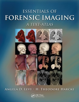
Bringing the long tradition of radiologic pathologic correlation to forensic radiology and autopsy, this volume provides readers with a technical and interpretive foundation for applying modern cross-sectional imaging to forensic autopsy.
Medical and industrial imaging methods have come to be recognized as powerful tools for documentation and data collection in many nontraditional settings.
Covering both noninvasive and surgical treatment alternatives, Critical Limb Ischemia defines practical guidelines for a multidisciplinary approach to critical limb ischemia and follows a step-by-step description of the latest techniques.
Updated to reflect the latest scientific advances and technologies in the diagnosis and treatment of pleural diseases, this new Second Edition explores the structure and function of these diseases and malignancies, from tuberculosis and asbestos to pleurisy and pneumothorax.
While it has aided far many more than it has harmed, radiation is forever etched in the public's mind as an indiscriminate and particularly pernicious killer.
To improve efficiency and reduce administrative costs, healthcare providers, insurance companies, and governments are increasingly using integrated electronic health record (EHR) and picture archiving and communication systems (PACS) to manage patients' medical information.
The benchmark first edition of Forensic Radiology, published in 1998, was a milestone in the forensic community - a bestseller throughout the world and a standard reference for practitioners and educators alike.
Answering the need that exists for a single reference to address the practical issues of implementing Image-guided Radiation Therapy (IGRT) into prostate cancer treatment, Image-guided Radiation Therapy of Prostate Cancer provides clinicians with a solid understanding of this technology and, through in-depth illustrations and step-by-step guideline
Maintaining the first edition's unique parallel to the strategy used by pathologists and pulmonologists to arrive at a patient's diagnosis in daily practice, Diagnostic Pulmonary Pathology starts with the patient and their biopsy findings, directing the pathologist or clinician to the proper diagnosis.
An ideal text for radiation oncologists, hematologist-oncologists, and radiologists, Image-Guided Radiotherapy and Functional Imaging in Modern Lymphoma Management is the foremost source for information on the increasingly important subject of image guided radiation therapy (IGRT) and its crucial role in the clinical evolution of high-precision ion
The Ischemic Penumbra presents the current status of concepts and research on this topic and identifies the latest methods for clinicians to quickly and efficiently recognize viable cerebral tissue for enhanced stroke management.
With the emergence of genetically manipulated laboratory mice as one of the most powerful tools for neuroscientists, imaging techniques capable of providing anatomical and functional information of small animals have become extremely important.
Kinematic MRI refers to imaging a joint through a range of motion to examine the interactions between the soft tissue and osseous anatomy that comprise the joint.
Image registration is the process of systematically placing separate images in a common frame of reference so that the information they contain can be optimally integrated or compared.
Magnetic Resonance Procedures: Health Effects and Safety is the first authoritative text on MR procedures and its associated health and safety concerns written by noted radiologists, physicists, and scientists with expertise in the field.
Vulnerable plaque development is the result of a complex series of molecular and cellular events involving inflammation, apoptosis, rupture, and thrombosis.
Providing a clear foundation as to what a clot is, how it forms, and the most recent approaches to treatment, Thrombus and Stroke is an all-inclusive resource covering: The fundamental science of clot formation and the pharmacokinetics of thrombolysis The clinical impact of thrombus as it pertains to stroke and the most recent clinical and minimally-invasive approaches to this disorder Current pharmacological interventions and the latest findings on neurovascular disease Clinical management of stroke Thrombogenicity of vascular implants In vivo stroke modeling Methods to assess the efficacy of anticoagulation therapy From the latest imaging techniques to endovascular treatments, this comprehensive, advanced volume provides critical data needed for diagnosis and treatment.
Musculoskeletal Radiology is a single-source guide encompassing all of musculoskeletal imaging, examining classical diseases, as well as modern interpretations of disease.
New Techniques in Cardiothoracic Imaging emphasizes emerging methods in computed tomography, magnetic resonance imaging, positron-emission tomography, and similar technology.
MRI-Guided Focused Ultrasound Surgery will be the first publication on this new technology, and will present a variety of current and future clinical applications in tumor ablation treatment.
The only source to fully cover every aspect of brain embolism, this guide analyzes the causes, symptoms, diagnosis, and management of this disorder-providing a detailed overview of major topics pertinent to embolism including donor sources, recipient sites, embolic material, recipient brain supply arteries, vascular and brain pathology, and the tre
Offering lower toxicity and higher accuracy than conventional therapies, this source offers illustrative coverage of this new method to treat tumors associated with brain, breast, lung, and neuroendocrine cancers.
This comprehensive reference discusses the use of ultrasound, computed tomography (CT), magnetic resonance imaging (MRI), and radioisotopes in the imaging of the adult and pediatric urinary tract.
With 2300 radiological images dispersed throughout the text, this source provides an expansive armamentarium of case studies and examples showcasing both common and uncommon liver pathologies.
This reference provides an authoritative overview of the role of ultrasonography and MR imaging technologies in the examination and assessment of the central nervous system of the fetus and neonate.
Exploring a technology that is significantly impacting the noninvasive evaluation of the physiology and anatomy of tumors, as well as the diagnosis of infectious processes and cardiac diseases, this source presents recent advances and clinical applications of sequential, single session single-photon emission computed tomography and computed tomography imaging.
This reference documents state-of-the-art trends and advancements in the utilization imaging modalities for the analysis of bones and their surrounding soft tissues, including muscles, tendons, ligaments, nerves, and blood vessels.
The ability of molecular and cellular imaging to track the survival, migration, and differentiation of cells in vivo as well as monitor particular gene expression in living subjects is rapidly moving from the research laboratory into daily clinical settings.
Although there are numerous books on drug metabolism, Radiotracers in Drug Development is unique in explaining how radiotracers are used to elucidate a drug's absorption, distribution, metabolism, and excretion (ADME).
In this comprehensive book, the author describes in a didactic and concise manner the sometimes misunderstood post-surgical aesthetic radiological aspects that can be misleading.
Gastrointestinal endoscopy is now mainstream and the focus is now changing from developing new techniques to enhancing the efficiency and quality of fundamental techniques.
This brand new text, is an essential practical guide for junior doctors and medical students making the transition from medical school to life on the wards.