Even the earliest applications of nuclear magnetic resonance (NMR) spectroscopy and tomography to medical inquiries, using experimental apparatus that was primitive by today's standards, demonstrated the extraordinary potential of the NMR method.
Radiology Today 3 presents papers and panel discussions from a multi- national faculty at the biannual Salzburg Symposium, which this time covered three important topics: critical diagnostic pathways in gastro"e; intestinal and genitourinary radiology, interventional radiology of the abdomen, and cost containment in radiology.
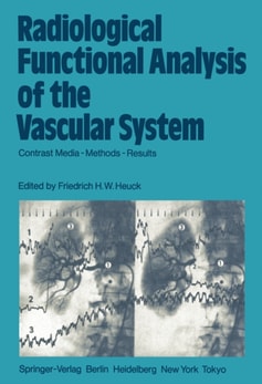
The quantitative analysis of blood flow within central and peripheral blood vessels has attracted more and more interest, for with the rapid developments in vascular surgery and the introduction of percutaneous transluminal angioplasty, it is becom- ing increasingly important to be able to measure regional blood flow in man.
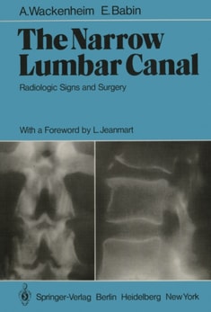
This book is intended for beginners and for those who want to refresh their knowledge of the elementary radioanatomy of the vertebrae, particularly their pathological radioanatomy.
Journalists, always very direct and in search of sensation, essentially asked me two questions on the occasion of this workshop: What were the goals of the meeting?
In this age when we are witnessing a veritable explosion in new modalities in diagnos- tic imaging we continue to have a great need for detailed studies of the vascularity of the brain in patients who have all types of cerebral vascular disease.
The purpose of this book is to provide the radiologist with information which is "e;as practical as possible"e; for the everyday use of computerized tomography (CT) in the field of cervical, thoracic, and musculoskeletal pathology.
It was our aim to place at the disposal of radiologists within a short time an atlas of high-quality, valuable pictures of abdominal CT without We felt that, the image degradation inherent to slower scanning apparatus.
More than 40 years ago British and German neurosurgeons met in Berlin and Breslau to exchange their experiences, to strengthen their friendly bonds, and to enjoy the attractions of both cities and their surroundings.
serves as an introduction to the anatomic-radiologic The use of whole-body axial transverse tomo- graphy (Scanner) as a new radio diagnostic method, data.
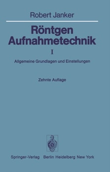
Das Buch zeigt Röntgenaufnahmen, die entsprechend den Einstellbeschreibungen in der "Röntgenaufnahmetechnik, Allgemeine Grundlagen und Einstellungen"* herge stellt wurden.
From the reviews: "e;Serre's notes on groups acting on trees have appeared in various forms (all in French) over the past ten years and they have had a profound influence on the development of many areas, for example, the theory of ends of discrete groups.
Bone and joint tuberculosis is common in developing countries, and surgeons in these countries are often faced with the dual problem of diagnosing and treating this disease.