The American Cancer Society anticipates that during 1995, 244000 new cases of cancer of the prostate will have been diagnosed in the United States, with 40400 deaths from the disease process.
Hyperthermia has been found to be of great benefit in combination with radiation therapy or chemotherapy in the management of patients with difficult and com- plicated tumor problems.
One of the most amazing and spectacular developments in modern radiology has been the rapid growth and expansion of so-called interventional radiology, which can also be described as minimally invasive therapy guided by radiological imaging.
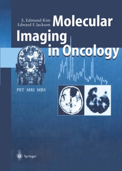
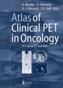
PET in Oncology describes the principles of positron emission tomography and is a useful resource for incorporating the technique in clinical practice.
Imaging studies are playing an increasingly role in the evaluation of endocrine diseases; accordingly, familiarity with the specific indications for the various modalities, and with the characteristic findings, is essential.
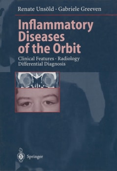
Numerous diseases in the areas of orthopaedics, rheumatology, and radiology can only be completely diagnosed if the corresponding conditions of the skin and mucous membranes are included in the diagnostic work-up (skin-bone).
In the United States in 1997, 28800 new cases of malignant tumors of the kidney and renal pelvis were diagnosed along with 2100 new cases of malignant tumors of the ureter and other urinary organs, 7200 primary malignant tumors of the testis, and 1300 primary malignant tumors of the penis and other genital organs.
Das Buch bietet Ärzten und Juristen vielseitige Hilfen zur Erstellung von Gutachten auf allen Fachgebieten der Medizin, die als Fundament für eine Rechtsprechung geeignet sind.
Diagnose und Therapie von Durchblutungsstörungen bei Veränderungen der Arterien und Venen sind mit der Angiographie erkennbar, und ihre Behandlung ist planbar geworden.
Although the technology required for the successful application of contrast echo cardiography has evolved rapidly over the past few years, the technique has not yet gained widespread clinical acceptance.
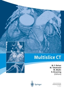
The introduction of multidetector spiral CT into clinical practice is without any doubt one of the most important technical developments in the field of computed tomography in general, and spiral CT in particular, in recent years.
Notwithstanding the important role of direct clinical and endoscopic examination in modern management of pathological conditions of the larynx, radiological study the and, more specifically, cross-sectional imaging by CT and MRI make definite diagnostic contributions by virtue of their potential to display superbly the deeper extent of laryngeal lesions.
This volume provides a multimodality imaging approach to the pathology of the foot and the ankle, covering both the various imaging techniques and all clinical aspects of diseases of these anatomic areas.
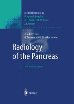
The pathology of the pancreas, which was a "e;hidden"e; retroperitoneal organ for many years, can now be marvelously depicted by the modern sectional imaging techniques.
Radiology of the Pancreas discusses the diagnostic role of the various imaging modalities currently available for the assessment of pancreatic anatomy and disease.
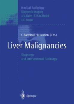
This book, written by leading experts from throughout the world, provides a comprehensive and up-to-date overview of the role of diagnostic and interventional radiology in respect of liver malignancies.
Hyperthermia has been found to be of great benefit in combination with radiation therapy or chemotherapy in the management of patients with difficult and com- plicated tumor problems.