In dem Nachschlagewerk wird ein Überblick über den Stellenwert verschiedener bildgebender Verfahren zur Differentialdiagnostik bestimmter Krankheitsbilder gegeben.
During recent years important progress in intensive medicine methods has fundamentally changed the approach to and the management of acute chest trauma, which in developed countries is frequently related to road accidents, the number of which is still rising.
Die neu überarbeitete und erweiterte Röntgenfibel dient dem angehenden Radiologen als Einführung und Hilfestellung in den verschiedenen Disziplinen seines Fachgebiets.
Although astonishing progress has been achieved during recent years in the treatment of patients afflicted with AIDS, this disease is still spreading in much of the developing world, where economic resources are limited.
The accurate diagnosis of tumors and tumor-like lesions of bones and soft tissues is often challenging, owing in part to the large number of such lesions and their shared clinical manifestations.
I have not embarked on the foreword to this scientific monograph by Armando and Gior- gio ROSSI in the expectation that it will be an easy task, because these two authors are the last remaining members of a family that has left its mark in the field of radiology in our country: therefore, the writer's enthusiasm and detachment could be jeopardized by mem- ories of his own teachers and elders and the respect he still feels towards them.
Important progress has been achieved during the past decade in the radiological diagnosis of congenital and acquired diseases of the kidneys and the urinary tract in infants and children.
Against the background of AIDS, wars, migration, and the inadequate provision of health care, a very serious epidemic of tuberculosis is once again occurring worldwide.
The numerous ways in which man and animals are affected by their physical environment, and the inborn and adaptive responses to change in the "e;milieu exterieur"e; have fascinated curious minds since the earliest days of recorded history.
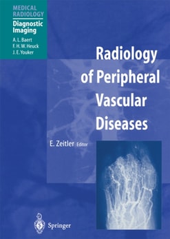
Based on the 1st edition this 2nd edition volume provides a completely revised comprehensive overview of the current state of the development in magnetic resonance (MR) vascular imaging.
Whereas during the past decade endoscopy has become established as the leading means of diagnosis and management of diseases affecting the esophagus, starnach and large bowel, radiology has retained its pre-eminence for the clinical study and evaluation of the small bowel.
Sonography, by virtue of its noninvasive nature, is used more and more in modern medicine as the first imaging modality for adults and children presenting with unex- plained cervical mass lesions as well as for the study of the carotid arteries and jugular veins.
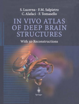
In the first half of the twentieth century, the study of neuroanatomy was essentiallybased on the observations made by scientists on brain cadavers fixed with standard techniques.
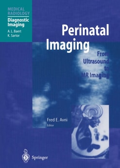
Fetal and perinatal medicine is a rapidly expanding field, and noninvasive imaging by means of ultrasonography and MRI is playing a major role in refining diagnosis and therapy.
The American Cancer Society anticipates that 16,500 patients will be diagnosed with primary malignant tumors of the central nervous system in 2000, with about 200,000 individuals presenting with brain metastases.
Since the publication of the first edition of Radiotherapy of Intraocular and Orbital Tumors in 1993 the treatment programs for cure have changed from the dominance of surgical resection to the utilization of radiation therapy with preservation of the eye intact and preservation of vision.
In dem vorliegenden Werk wird erstmals für den deutschsprachigen Raum ein Überblick gegeben über: die theoretischen Voraussetzungen einer regionalen Therapie, die verschiedenen Techniken zu deren Durchführung und über die bisher gewonnenen Ergebnisse unter Berücksichtigung des jeweiligen Tumortyps, seiner Lokalisation und der angewandten Form der regionalen Therapie.
Das Buch vermittelt dem Radiologen wertvolle Hinweise auf die klinischen Zusammenhänge und gibt auf der anderen Seite dem Kliniker Einblick in die vielfältigen Möglichkeiten der kernspintomographischen Diagnostik.
Our goal for this second edition of Oncologic Therapies is to again provide a brief introduction to the principles and practice of oncology and to oncologic therapies.
Mammography can be thought of as a (sub)macroscopic "e;shadow pathology"e; - confident diagnosis necessitates comprehensive knowledge of breast pathology.
This book describes and summarizes the radiation responses of both normal and neoplastic tissues with a focus on rational strategies for the modification of these responses.
Nuclear Medicine techniques have advanced to such a degree that biochemical transparency of the human body has reached the doorstep of medical application.
Eine gelungene, aussagekräftige Ultraschalluntersuchung erfordert zweierlei: Ebenso wichtig wie ein technisch guter Schallkopf ist ein in der sonographischen Methode gut geschulter Untersucher.