Detection and characterization of bone tumors with imaging remains a big challenge for every radiologist notwithstanding the impressive progress achieved by the introduction of several new imaging modalities.
It is a great privilege to introduce this book devoted to the current and future roles in research and clinical practice of another exciting new development in MRI: Diffusi- weighted MR imaging.
Radiation Oncology: An Evidence-Based Approach (ROEBA) is a reference book designed to enable radiation oncologists, including those in training, to make diagnostic and treatment decisions on the basis of the best available scientific evidence.
Radioisotope-based molecular imaging probes provide unprecedented insight into biochemistry and function involved in both normal and disease states of living systems, with unbiased in vivo measurement of regional radiotracer activities offering very high specificity and sensitivity.
In this book every urologic procedure is described in a step by step sequence of events and the text is supplemented with innumerous Tips, colored illustrations and high definition photographs depicting the main steps of the procedures.
Te rapidly changing concepts in radiation oncology with the development of more precise - strumentation for delivery of radiation therapy and a greater emphasis on hypofractionation technologies require a very intimate knowledge of tumor biology and the infuence of various biologic factors on dose distribution within the tumor in terms of homogeneity as well as prev- tion of any late efects on normal tissue surrounding the tumor itself.
Based on the experience gained by PET/CT experts with more than 10,000 patients, this manual impressively demonstrates the advantages of combined PET/CT.
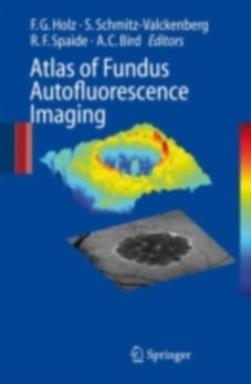
During recent years, FAF (Fundus autofluorescence) imaging has been shown to be useful in various retinal diseases with regard to diagnostics, documentation of changes, identification of disease progression, and monitoring of novel therapies.
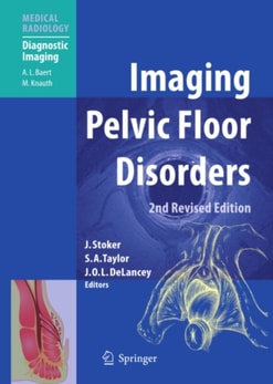
This volume builds on the success of the first edition of Imaging Pelvic Floor Disorders and is aimed at those practitioners with an interest in the imaging, diagnosis and treatment of pelvic floor dysfunction.
Dural cavernous sinus fistulas (DCSFs) are benign vascular diseases consisting in an arteriovenous shunt at the cavernous sinus that if misdiagnosed can lead to potentially serious ophthalmologic complications.
Parallel imaging techniques have only recently been introduced into magnetic resonance imaging (MRI) in clinical routine, but they have already gained wide clinical acceptance in numerous applications.
Because of the radiation dose delivered, multidetector row CT (MDCT) may induce cancers, and the risk of death has been estimated at up to one per 1,000 examinations.
In the Name of Love and Family This book could not have been completed without the continuing support of my wife, Isabelle, who, as a radiologist herself, not only understood my end- vours to complete this work, but who was also my most loyal supporter.
In March 1973, a short paper was published in Nature, entitled Image formation by induced local int- action; examples employing nuclear magnetic resonance.