Imaging of the temporal bone has recently been advanced with multidetector CT and high-field MR imaging to the point where radiologists and clinicians must familiarize themselves with anatomy that was previously not resolvable on older generation scanners.
Over the past decade, PET-CT has achieved great success owing to its ability to simultaneously image structure and function, and show how the two are related.
Since the publication of the first edition of Nuclear Hepatology almost 10 years ago, new advances have taken place, both in our understanding of liver pathophysiology and various imaging modalities.
This handy, well-illustrated manual has been designed to meet the need of interventional pain physicians to understand the radiologic imaging involved in the performance of non-spinal pain procedures.
Nasopharyngeal Cancer - Multidisciplinary Management provides a comprehensive account of the current state of knowledge on nasopharyngeal cancer and its multidisciplinary management.
A general consensus exists,that lumbosacral nerve root compression is the primary cause of sciatica and neurogenic claudication, although humoral and vascular factors certainly play a role as well.
Although [18F]fluorodeoxyglucose (FDG) generally shows an excellent performance as a cancer-imaging agent when using PET-CT, there are some settings in which other radiopharmaceuticals offer advantages.
This book is an ideal introduction to the use of radiology in imaging diseases of the liver, gallbladder and biliary system, pancreas, spleen, and gastrointestinal tract.
Functional magnetic resonance imaging (fMRI) and Electronecephalography (EEG) are very important and complementary modalities since fMRI offers high spatial resolution and EEG is a direct measurement of neuronal activity with high temporal resolution.
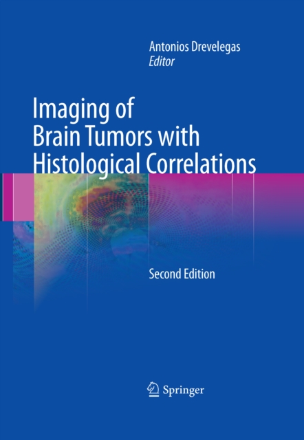
This volume provides a deeper understanding of the diagnosis of brain tumors by correlating radiographic imaging features with the underlying pathological abnormalities.
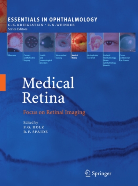
Recent advances in ophthalmic imaging technology have revolutionized fundus examination and contributed significantly in elucidating the pathophysiology of retinal diseases and improving their diagnosis and treatment.
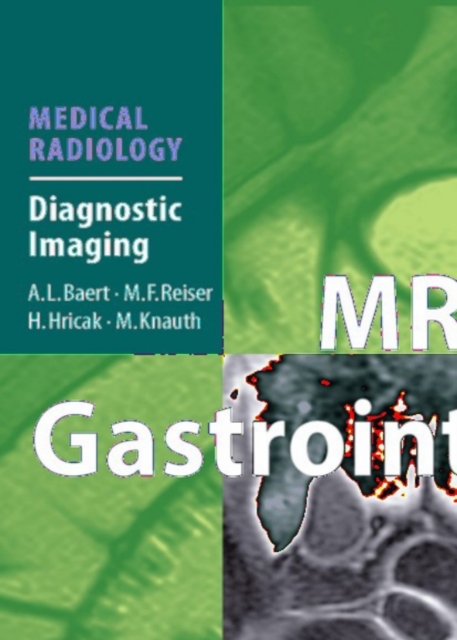
MRI has become an important tool in the management of patients with diseases of the gastrointestinal tract, such as rectal cancer and inflammatory bowel diseases.
This book adopts a multidisciplinary approach to examine in detail a range of interesting new concepts in the diagnosis and therapy of pancreatic adenocarcinoma.
This book describes the current status of the very rapidly developing field of high-field MR and examines the possibilities, challenges, and limitations of this fascinating technology.
Neural activity in the human brain generates coherent synaptic and intracellular currents in cortical columns that create electromagnetic signals which can be measured outside the head using magnetoencephalography (MEG) and electroencephalography (EEG).
Rapid progress in the technique and practice of virtual colonoscopy as well as the conti- ing clinical high interest for this radiodiagnostic procedure made this second edition, only 3 years after the publication of the ?
Nuclear medicine technology has been a fascinating subject ever since the early rectilinear scanner produced images, and is becoming ever more interesting (and demanding) with the growth of positron emission tomography and other molecular imaging.
Keywords Spin Electromagnetic radiation Resonance Nucleus Hydrogen Proton Certain atomic nuclei possess inherent magnetic Let us summarize the MRI procedure.
Detection and characterization of bone tumors with imaging remains a big challenge for every radiologist notwithstanding the impressive progress achieved by the introduction of several new imaging modalities.
It is a great privilege to introduce this book devoted to the current and future roles in research and clinical practice of another exciting new development in MRI: Diffusi- weighted MR imaging.
Radiation Oncology: An Evidence-Based Approach (ROEBA) is a reference book designed to enable radiation oncologists, including those in training, to make diagnostic and treatment decisions on the basis of the best available scientific evidence.
Radioisotope-based molecular imaging probes provide unprecedented insight into biochemistry and function involved in both normal and disease states of living systems, with unbiased in vivo measurement of regional radiotracer activities offering very high specificity and sensitivity.