Intravascular ultrasound (IVUS) imaging has become an essential tool for understanding arterial disease and guiding percutaneous interventions in the contemporary era.
While there are many excellent texts focused on clinical medical imaging, there are few books that approach in vivo imaging technologies from the perspective of a scientist or physician-scientist using, or interested in using, these techniques in research.
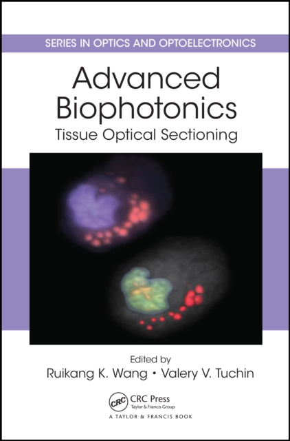
Despite a number of books on biophotonics imaging for medical diagnostics and therapy, the field still lacks a comprehensive imaging book that describes state-of-the-art biophotonics imaging approaches intensively developed in recent years.
Renowned as the essential reference for all venous specialists, phlebologists, vascular surgeons, and vascular medicine physicians, the fifth edition of the Handbook of Venous and Lymphatic Disorders: Guidelines of the American Venous Forum covers the investigation, prevention, evaluation, and treatment of venous and lymphatic disorders.
This textbook, offers a quick and easy introduction to the fundamentals of hip sonography as standardised screening tool for detecting developmental dysplasia and dislocation of the hip (DDH), thus allowing for early treatment in the neonatal period.
This textbook, offers a quick and easy introduction to the fundamentals of hip sonography as standardised screening tool for detecting developmental dysplasia and dislocation of the hip (DDH), thus allowing for early treatment in the neonatal period.
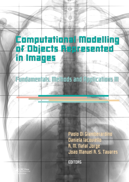
Computational Modelling of Objects Represented in Images: Fundamentals, Methods and Applications III contains all contributions presented at the International Symposium CompIMAGE 2012 - Computational Modelling of Object Presented in Images: Fundamentals, Methods and Applications (Rome, Italy, 5-7 September 2012).
Image Processing with MATLAB: Applications in Medicine and Biology explains complex, theory-laden topics in image processing through examples and MATLAB algorithms.
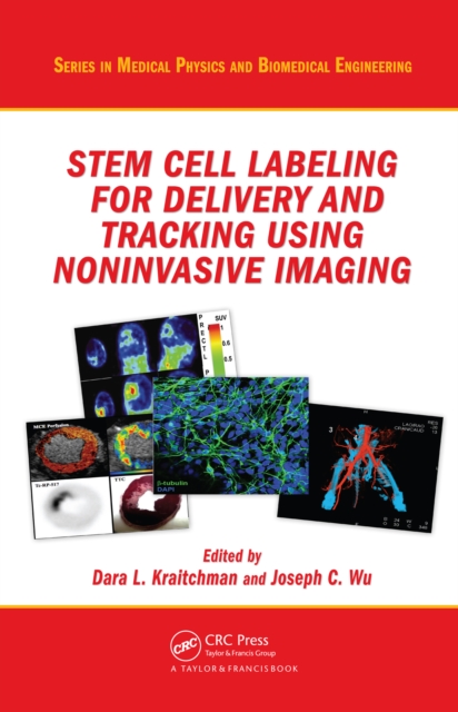
Stem Cell Labeling for Delivery and Tracking Using Noninvasive Imaging provides a comprehensive overview of cell therapy imaging, ranging from the basic biology of cell therapeutic choices to the preclinical and clinical applications of cell therapy.
This book provides evidence-based guidance for pediatric imaging to radiologists, pediatricians, and all subspecialists caring for children, as well as policy advocates.
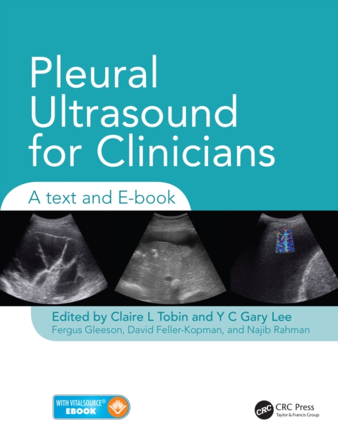
With a large volume of ultrasound images and teaching videos, Pleural Ultrasound for Clinicians: A Text and E-book is a print and interactive digital resource that provides practical guidance for all those undertaking pleural ultrasound investigations and procedures.
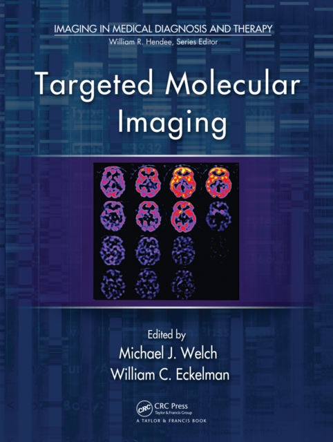
Targeted Molecular Imaging covers the development of novel diagnostic approaches that use an imaging probe and agent to noninvasively visualize cellular processes in normal and disease states.
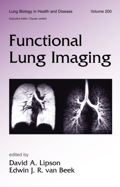
Written by world authorities, this reference explores the utilization and advancement of state-of-the-art imaging modalities for the analysis of pulmonary function-providing an up-to-date and global perspective of imaging applications with detailed contributions from experts in Europe, North America, Japan, and Australia.
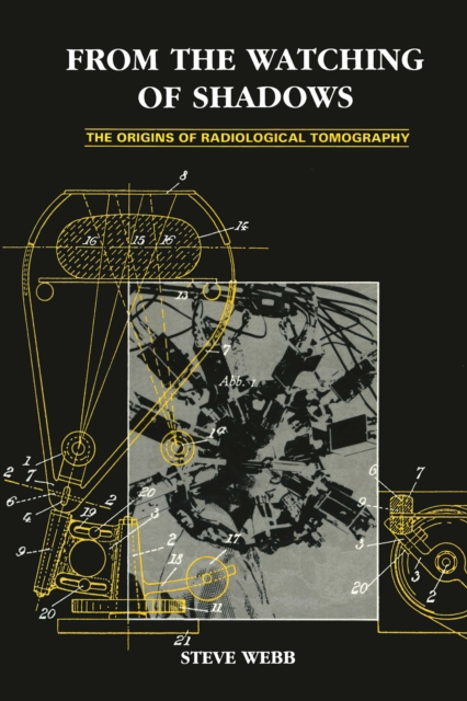
From the Watching of Shadows: The Origins of Radiological Tomography presents the first complete history of body imaging by discrete sections, from its earliest beginnings around 1920 to modern times.
Physiological optical imaging is a group of emerging technologies that aim to provide healthcare practitioners and biomedical researchers with information about tissue physiology or pathophysiology using approaches different from traditional medical imaging (PET, ultrasound, MRI, X-ray, or CT scan).
The Ischemic Penumbra presents the current status of concepts and research on this topic and identifies the latest methods for clinicians to quickly and efficiently recognize viable cerebral tissue for enhanced stroke management.
From the Lab to Clinical Settings-Advances in Quantitative, Noninvasive Optical DiagnosticsNoninvasive fluorescence imaging techniques, novel fluorescent labels, and natural biomarkers are revolutionizing our knowledge of cellular processes, signaling and metabolic pathways, the underlying mechanisms for health problems, and the identification of n
Completely up to date with the latest examination changes, Get Through First FRCR: MCQs for the Physics Module offers a valuable insight into the new Physics module of the First FRCR examination.
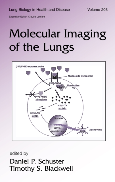
This authoritative reference reviews current and emerging molecular imaging modalities that are expected to impact the detection and treatment of lung diseases in the near future-including applications in gene expression and inflammation imaging; the imaging of pulmonary cytokine regulation; the molecular imaging of angiogenesis, the airways, and l
Adaptive Optics (AO) is a technique allowing the manipulation of the wavefront of the light benefitting multiple fields like Microscopy, Optical Coherence Tomography, Visual Testing, Optical Communications, and Astronomical Optics, just to mention some examples.
This book examines Dynamic Light Scattering (DLS) and its derivatives Laser Doppler Flowmetry (LDF), Diffusing Wave Spectroscopy (DWS), Laser Speckle Contrast Imaging (LSCI), and Doppler Optical Coherence Tomography (OCT) for characterizing particle motion in turbid mediums like suspensions and solutions.
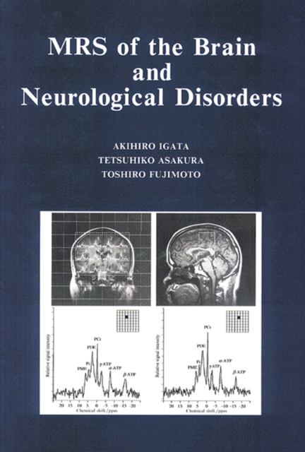
MRS of the Brain and Neurological Disorders illustrates and demonstrates the usefulness of magnetic resonance spectroscopy for the diagnosis of brain and neurological disorders, including epilepsy, nervous disorders, psychiatric disorders, and brain tumors.
Written by skilled specialists in the field of interventional pulmonology, the new Second Edition thoroughly explores the latest advancements, newest therapies, and diagnostic techniques in interventional pulmonary medicine.
This text focuses on the anatomical, physiological, pathological, and technical aspects of post robotic prostatectomy and post radiation urinary continence and erectile function.
This text focuses on the anatomical, physiological, pathological, and technical aspects of post robotic prostatectomy and post radiation urinary continence and erectile function.
In regenerative medicine, tissue engineers largely rely on destructive and time-consuming techniques that do not allow in situ and spatial monitoring of tissue growth.
Second-harmonic generation (SHG) microscopy has shown great promise for imaging live cells and tissues, with applications in basic science, medical research, and tissue engineering.
"e;Biomedical Imaging: Principles and Advancements"e; offers a captivating exploration of the intricate landscapes within the human body, revealing the transformative power of biomedical imaging.