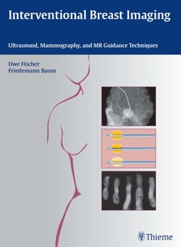
A step-by-step guide to performing image-guided breast interventionsWritten by a team of experts, this practical how-to guide provides a systematic overview of the most current image-guided interventional techniques used to diagnose breast abnormalities.
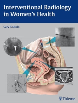
A comprehensive reference for optimizing care in interventional radiology for womenThis book is a practical resource for the current applications of interventional radiology in the care of female patients.
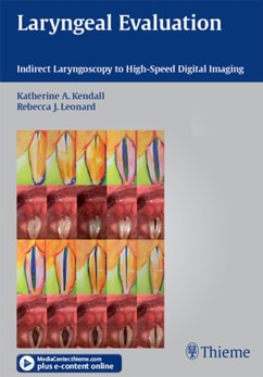
A must-have multimedia reference on the latest laryngeal examination and imaging techniquesThis comprehensive, full-color reference provides a thorough overview of the most recent advances in laryngeal imaging technology combined with all of the information readers need to interpret findings and successfully manage patients with voice disorders.
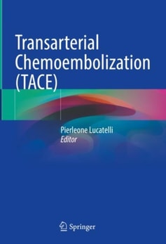
The book thoroughly presents Transarterial Chemoembolization (TACE), a procedure representing the standard of care for several clinical oncological indications.
Expert guidance on managing interventional radiology in traumaInterventional Radiology in Trauma Management brings together the insights and expertise of Dr.
X-ray computed tomography (CT) has been one of the most popular diagnostic imaging modalities for decades in the clinic for saving patients' lives or improving their quality of life.
This book provides an overview on the critical role of diagnostic imaging in the assessment of patients with suspected alimentary tract perforation, an emergent condition that requires prompt surgery.
This book, featuring more than 180 high spatial resolution images obtained with state-of-the-art MDCT and MRI scanners, depicts in superb detail the anatomy of the temporal bone, recognized to be one of the most complex anatomic areas.
This book provides a detailed and comprehensive overview of the role of diagnostic imaging in the assessment and management of trauma and polytrauma in children.
The book provides a comprehensive description of the basic ultrasound principles, normal anatomy of the lower limb muscles and classification of muscle strain injuries.
With contributions from an esteemed otolaryngologist, talented photographer, and multidisciplinary specialists, Mastering Medical Photography of the Head and Neck demystifies the process of medical photography.
A practical imaging primer designed specifically for ENTsImaging for Otolaryngologists distils the essentials of otolaryngologic imaging into a concise reference that concentrates on key topics that are of immediate interest to otolaryngologists practicing in a modern clinical environment.
Unique case-based reference presents high-yield images and expertise focused on vascular neuroradiologyImaging in Neurovascular Disease: A Case-Based Approach by Waleed Brinjikji and Timo Krings is unique in its approach, detailing diagnostic and interventional neuroradiology cases based on radiologic findings.
This handbook is a concise practical guide for residents and general radiologists that will offer reliable assistance during the performance and reporting of multidetector row computed tomography and magnetic resonance imaging in patients with Liver Bile Ducts and Pancreas conditions.
In recent years, there has been huge interest in developing new methods that offer improved accuracy for the detection of small bowel pathology, and in particular for the assessment of inflammatory bowel diseases (IBD).
This new second edition presents a completely new selection of 25 scenarios based on cases from the personal archive of Ondrej Dolezal, collected over 20 years of clinical practice.
A new third edition of the outstanding introduction to radiologic imagingAs an overview to radiology this high quality text from Thieme provides a comprehensive picture of current imaging practice and is suitable for reading by a range of healthcare professionals at undergraduate or post-graduate level.
Authoritative and lavishly illustrated, this best-selling reference returns in a fourth edition with comprehensive coverage of the current imaging strategies for the evaluation of disease processes affecting the temporal bone and its intricate anatomy.
This book offers the first non-official history of French nuclear policies which goes beyond the divide between nuclear weapons and nuclear energy policies.
This book offers the first non-official history of French nuclear policies which goes beyond the divide between nuclear weapons and nuclear energy policies.
This book on the anatomy of central nervous system arteries concentrates on all anatomical variations of the central nervous system and it describes the embryological processes that hide behind the possible adult variants.
This book comprehensively covers the diagnosis, classification, assessment, and management of shoulder arthritis, a condition with increasing incidence that affects people of all ages.
This book includes detailed discussions of the latest science in the embryogenesis of spinal dysraphic malformations, and a well-illustrated guide to their surgical repair.
Digital Breast Tomosynthesis: Technique and Cases is a comprehensive and timely introduction to a significant technological advance in breast cancer imaging.
This book includes detailed discussions of the latest science in the embryogenesis of spinal dysraphic malformations, and a well-illustrated guide to their surgical repair.
FIVE STARS from Doody's Star RatingsAuthored by renowned UEFA specialists in the medical care of football players, this three-volume series-sourced from the course materials used in UEFA's Football Doctor Education Program-aims to familiarize clinicians with a structured system of assessment and care in dealing with the wide variety of injuries that can afflict professional footballers.
The definitive guide to MRI for musculoskeletal abnormalitiesDifferential Diagnosis in Musculoskeletal MRI is a unique desk reference offering extensive descriptions of MRI findings that enable radiologists to more easily diagnose a wide range of musculoskeletal conditions.
This book, written by internationally renowned experts, is a comprehensive text covering all aspects and recommendations regarding the use of POCUS for critically ill neonates and children.
This handbook concisely summarizes state-of-the-art information about stereotactic radiosurgery (SRS) and stereotactic body radiotherapy (SBRT), including the history and development of these modalities, the biologic rationale for these technologies, typical practices, and reported results.
Your one-stop source of complete imaging information for the evaluation of thoracic conditions and diseases in all modalitiesDue to the remarkable concentration of various vital organs that can be visualized in thoracic imaging, the region occupies a firm central place in the spectrum of diagnostic imaging.