Ultrasound in Medicine is a broad-ranging study of medical ultrasound, including ultrasound propagation, interaction with tissue, and innovations in the application of ultrasound in medicine.
Charred, badly decomposed, or mummified corpses, as well as those restrictions forced upon coroners by certain religious sects, often make autopsies impossible to perform.
Edited by and featuring contributions from world-class researchers, Ophthalmological Imaging and Applications offers a unified work of the latest human eye imaging and modeling techniques that have been proposed and applied to the diagnosis of ophthalmologic problems, including inflammation, cataracts, diabetic retinopathy, and glaucoma.
Cardiovascular Magnetic Resonance (CMR) is well established in clinical practice for the diagnosis and management of a wide array of cardiovascular diseases.
Shaped by Quantum Theory, Technology, and the Genomics RevolutionThe integration of photonics, electronics, biomaterials, and nanotechnology holds great promise for the future of medicine.
The evolution of technological advances in infrared sensor technology, image processing, "e;smart"e; algorithms, knowledge-based databases, and their overall system integration has resulted in new methods of research and use in medical infrared imaging.
The field of magnetic resonance imaging (MRI) has developed rapidly over the past decade, benefiting greatly from the newly developed framework of compressed sensing and its ability to drastically reduce MRI scan times.
Illustrating the development and determinants of possible plaque rupture, Cardiovascular Plaque Rupture provides an in-depth review of the pathology, etiology, cellular and molecular biology, diagnosis, and treatment of plaque in the cardiovascular system-exploring the roles of physical stress, inflammation, and infectious agents on the growth of c
An innovative, three-dimensional x-ray imaging technique that enhances projection radiography by adding depth resolution, Tomosynthesis Imaging explores tomosynthesis, an emerging limited-angle tomographic imaging technology that is being considered for use in a range of clinical applications, and is currently being used for breast cancer screening
As the third leading cause of death in the United States, stroke accounts for one in every fifteen deaths and is the major cause of disability in the country.
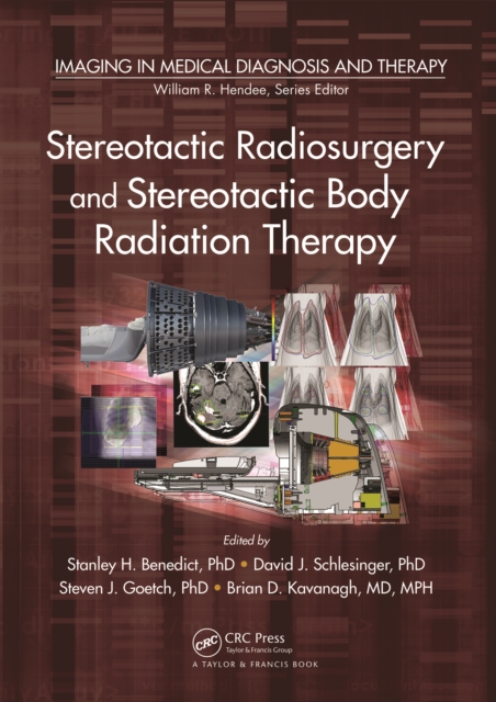
Written by internationally known experts in the field, Stereotactic Radiosurgery and Stereotactic Body Radiation Therapy examines one of the fastest-developing subspecialties within radiation oncology.
Comprised of chapters carefully selected from CRCa s best-selling engineering handbooks, volumes in the Principles and Applications in Engineering series provide convenient, economical references sharply focused on particular engineering topics and subspecialties.
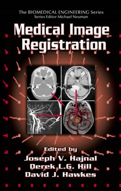
Image registration is the process of systematically placing separate images in a common frame of reference so that the information they contain can be optimally integrated or compared.
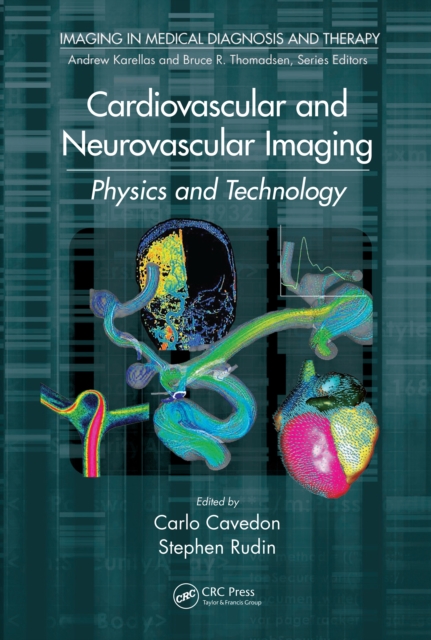
Cardiovascular and Neurovascular Imaging: Physics and Technology explains the underlying physical and technical principles behind a range of cardiovascular and neurovascular imaging modalities, including radiography, nuclear medicine, ultrasound, and magnetic resonance imaging (MRI).
Emphasizing new and emerging therapies in each chapter, this reference provides essential information for clinicians to provide accurate diagnoses and select the most appropriate treatment regimens for patients with primary and metastatic brain tumors and neurological complications of cancer.
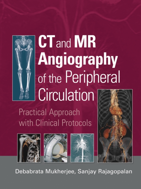
This text discusses the basic aspects of multislice CT angiography with chapters on technical principles, basic scan technique for peripheral vascular imaging with multislice CT, image reconstruction with multislice CT, radiation doses, and contrast agent administration.
This book is discussing artificial intelligence-based smart systems for diagnosis and prediction of diseases, artificial intelligence in clinical decision-making, and artificial intelligence tools for clinical and healthcare data.
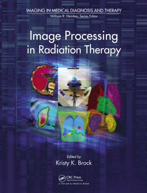
Images from CT, MRI, PET, and other medical instrumentation have become central to the radiotherapy process in the past two decades, thus requiring medical physicists, clinicians, dosimetrists, radiation therapists, and trainees to integrate and segment these images efficiently and accurately in a clinical environment.
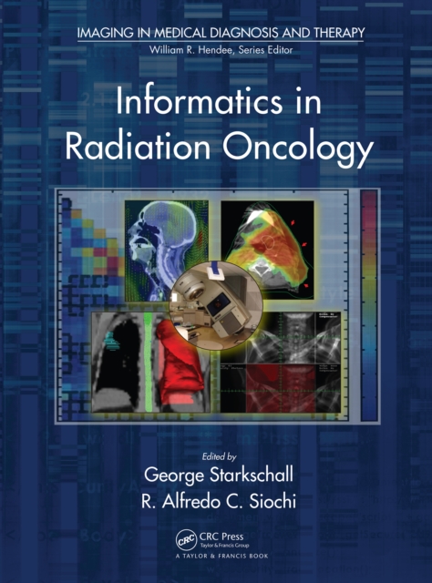
Reflecting the increased importance of the collaborations between radiation oncology and informatics professionals, Informatics in Radiation Oncology discusses the benefits of applying informatics principles to the processes within radiotherapy.
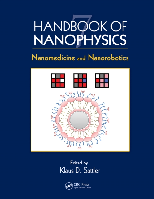
The tools of nanodiagnostics, nanotherapy, and nanorobotics are expected to revolutionize the future of medicine, leading to presymptomatic diagnosis of disease, highly effective targeted treatment therapy, and minimum side effects.
Rapid evolution of technical advances in infrared sensor technology, image processing, 'smart algorithms, databases, and system integration paves the way for new methods of research and use in medical infrared imaging.
At the cutting-edge of maternal and neonatal management and research, this reference presents the most recent advances in fetal DNA/RNA, embryoscopy/fetoscopy, open fetal surgery, and ethnic population screening to manage pre-existing and newly detected maternal medical complications, detect genetic and congenital abnormalities, prevent preterm bir
In considering ways that physics has helped advance biology and medicine, what typically comes to mind are the various tools used by researchers and clinicians.
Enhanced, more reliable, and better understood than in the past, artificial intelligence (AI) systems can make providing healthcare more accurate, affordable, accessible, consistent, and efficient.
Computational Optical Biomedical Spectroscopy and Imaging covers recent discoveries and research in the field by some of the best inventors and researchers in the world.
Edited by a renowned international expert in the field, Nuclear Medicine Physics offers an up-to-date, state-of-the-art account of the physics behind the theoretical foundation and applications of nuclear medicine.
The First Book on CRS MicroscopyCompared to conventional Raman microscopy, coherent Raman scattering (CRS) allows label-free imaging of living cells and tissues at video rate by enhancing the weak Raman signal through nonlinear excitation.
In the medical imaging field, clinicians and researchers are increasingly moving from the qualitative assessment of printed images to the quantitative evaluation of digital images since the quantitative techniques often improve diagnostic accuracy and complement clinical assessments by providing objective criteria.
Supported by an extensive review of migraine pathogenesis and more than 200 case histories gleaned from the authors' personal experiences, this guide proposes two radical new concepts: all primary headaches are fundamentally a form of migraine and everyone may experience some manifestation of the migraine process in their life.
Filled with an abundance of reader-friendly and quick-reference tables and figures, this Fourth Edition spans the etiology, epidemiology, clinical features, diagnosis, pathology, and treatment of multiple sclerosis (MS).
Written by an interdisciplinary team of medical doctors, computer scientists, physicists, engineers, and mathematicians, Correction Techniques in Emission Tomography presents various correction methods used in emission tomography to generate and enhance images.
This book is discussing artificial intelligence-based smart systems for diagnosis and prediction of diseases, artificial intelligence in clinical decision-making, and artificial intelligence tools for clinical and healthcare data.
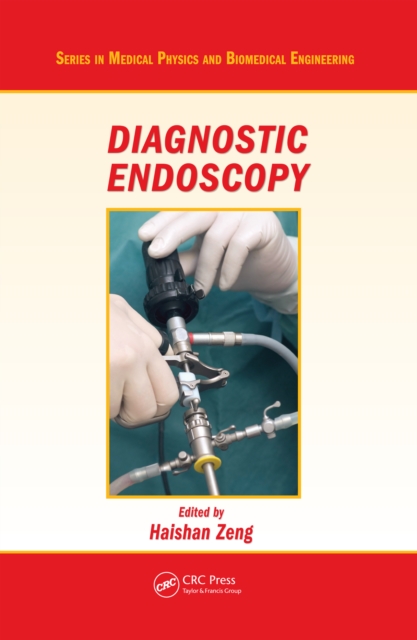
Combining a professional development course on diagnostic endoscopy from SPIE (the international society advancing light-based research) and the authors' graduate course on biomedical optics, this work is written for researchers in medical optics and biomedical engineering as well as graduate medical optics students.
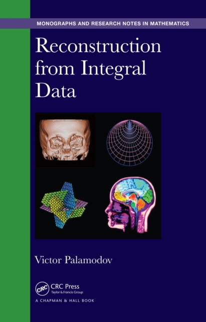
Reconstruction of a function from data of integrals is used for problems arising in diagnostics, including x-ray, positron radiography, ultrasound, scattering, sonar, seismic, impedance, wave tomography, crystallography, photo-thermo-acoustics, photoelastics, and strain tomography.
Discover the Most Advanced Technologies in BiomagneticsCo-edited by Professor Ueno, a leader in the biomagnetics field for over 40 years, Biomagnetics: Principles and Applications of Biomagnetic Stimulation and Imaging explains the physical principles of biomagnetic stimulation and imaging and explores applications of the latest techniques in neuroscience, clinical medicine, and healthcare.
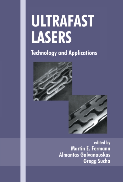
Covering high-energy ultrafast amplifiers and solid-state, fiber, and diode lasers, this reference examines recent developments in high-speed laser technology.
Providing a detailed study of the anatomy, physiology, and pathology of peripheral nerves, this reference provides a clear approach to the evaluation, characterization, and treatment of peripheral neuropathies-reviewing each major class of these disorders with authoritative discussions of clinical characteristics, electrodiagnostic features, examin
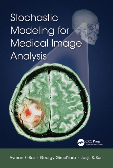
The field of medical imaging has been revolutionized by new techniques in powerful computations, image processing, and modalities such as Computer-Aided Tomography (CAT) and Magnetic Resonance Imaging (MRI), among others.
With over 100 illustrations, Volume 1 addresses the core disciplines of pharmaceutics (absorption, PK, excipients, tablet dosage forms, and packaging), and explores the challenges and paradigms of pharmaceutics.
From the discovery of x-rays in 1895 through the emergence of computed tomography (CT) in the 1970s and magnetic resonance imaging (MRI) in the 1980s, non-invasive imaging has revolutionized the practice of medicine.