As fMRI technology has provided invaluable insights into the mechanisms through which the human brain works in healthy individuals and in patients with different neurological and psychiatric conditions, the study of brain function and even the monitoring of the effects of treatment have become more effective and efficient.
The advent of non-invasive imaging technology, such as magnetic resonance imaging (MRI), has allowed biologists and clinicians to make great strides in unraveling the secrets of the brain.
This monograph, now in its 2nd edition with 31 new chapters and significant updates, is the first book of its kind written specifically for graduate students and clinicians.
This book highlights the integration of science and imaging and demonstrates how we can teach and learn in a much more accessible, innovative, and engaging way using technology.
This book describes the different aspects of ischemic heart disease, one of the leading causes of death in industrialized countries, and whose growing prevalence represents a major challenge in terms of short- and long-term management and surveillance.
Imaging in critically ill patients is a ubiquitous but challenging line of investigation for the physician as accurate interpretation is often difficult as patient cooperation during the procedure is grossly compromised, and the resultant image is often suboptimal.
Imaging in critically ill patients is a ubiquitous but challenging line of investigation for the physician as accurate interpretation is often difficult as patient cooperation during the procedure is grossly compromised, and the resultant image is often suboptimal.
This easy-to-understand pocketbook in the highly respected Clark's stable of imaging texts is an invaluable tool and training aid, providing essential information for mammographic positioning, technique and interpretation for mammography practitioners at all levels.
Technological innovations accompanying advances in medicine have given rise to the possibility of obtaining better-defined fetal images that assist in medical diagnosis and contribute toward genetic counseling offered to parents during the prenatal care.
Handbook for Clinical Trials of Imaging and Image-Guided Interventions is the first single-source, multi-disciplinary reference, based on the didactic sessions presented at the annual Clinical Trials Methodology Workshop for radiologists, radiation oncologists and imaging scientists (sponsored by the Radiological Society of North America (RSNA)).
This practical guide presents an up-to-date, comprehensive yet concise collection of the most frequently used measurements in musculoskeletal studies carried out in children and adolescents through the different radiological modalities.
This third edition offers comprehensive, up-to-date coverage of all areas of interventional pulmonology, a minimally invasive endoscopic method for diagnosing and treating lung disorders.
Improve your imaging interpretation skills for the most commonly encountered surgical conditionsThe goals of Acute Care Surgery: Imaging Essentials for Rapid Diagnosis is help acute care surgeons, general surgeons, and surgical trainees develop the skills necessary to efficiently work up and diagnose critical surgical disease.
This extensively updated edition provides a comprehensive review of intensive care for neurologically injured patients from the emergency room and ICU through the operating room and post-surgical period in two comprehensive volumes.
The most authoritative guide to sonography in obstetrics and gynecology-now in full colorCompanion website includes images, case studies, and more Written by radiologists and ob/gyns to provide a balanced perspective, this standard-setting guide is both a clinically relevant reference text and atlas presented in full color for the first time.
Imaging the Addicted Brain, the latest volume in the International Review of Neurobiology series will appeal to neuroscientists, clinicians, psychologists, physiologists, and pharmacologists.
A concise, outline-format review of the curriculum based on the ARDMS content outline More than 3,000 registry-format review questions and answers with complete explanations NEW chapters on 3D sonography in obstetrics and gynecology, musculoskeletal sonography, and breast sonography NEW: Now includes an index to make locating information seamless Fully referenced to core books students are likely to have on hand for further study
Market includes physical therapists, physical therapy and occupational therapy studentsState-of-the-art images illustrate the injury and healing processIncludes a suggested treatment section for each injury listedHighly visual: 330 illustrationsCovers radiography, CT, MRI, and ultrasound from the perspective of the therapist
Everything radiography students need to ace the certification examHailed by Doody s Review Service as the gold standard among instructors and students , Radiography PREP delivers a concise summary of the entire radiography curriculum in a readable narrative.
A comprehensive review for the mammography registry examination - from an experienced educator and clinician who knows exactly what it takes to passIncludes new coverage of the latest digital imaging technologies Written by an instructor and mammography specialist at Stamford HospitalConcise narrative text helps you to focus on essential concepts Practice questions with answers referenced to the text allow you to gauge your comprehension of important material Learning aids such as objectives and glossaries at the beginning of each chapter streamline the learning processNumerous radiographs teach you to recognize good and bad films and normal circumscribed lesions and breast calcifications High-quality diagrams help you learn correct patient positioning consistent with the American College of Radiography and the Mammography Quality Control Manual Valuable during coursework to help you recognize and understand concepts that are likely to appear on the exam A complete review for licensure that includes the history of breast imaging, breast cancer detection, and treatment (including new imaging methods and recent advances in digital mammography, MRI, BSGI, DBT, volumtetric ultrasound imaging, and Cone Beam Breast CT)
A complete introductory text to musculoskeletal imagingBasic Musculoskeletal Imaging is an engagingly written, comprehensive textbook that addresses the fundamental principles and techniques of general diagnostic and advanced musculoskeletal imaging.
366 cases and more than 3000 images help you accurately interpret head and neck imagingHead and Neck Imaging Cases uses 366 cases and more than 3000 images to familiarize you with imaging findings of common head and neck diseases and conditions encountered in daily practice.
A State-of-the-Art Guide to Biomedical Engineering and Design Fundamentals and ApplicationsThe two-volume Biomedical Engineering and Design Handbook, Second Edition offers unsurpassed coverage of the entire biomedical engineering field, including fundamental concepts, design and development processes, and applications.
With treatment approaches and the field of neuro-oncology neuroimaging changing rapidly, this third edition of the Handbook of Neuro-Oncology Neuroimaging is very relevant to those in the field, providing a single-source, comprehensive, reference handbook of the most up-to-date clinical and technical information regarding the application of neuroimaging techniques to brain tumor and neuro-oncology patients.
A dynamic, all-inclusive overview of the field of health physicsIf it's an important topic in the field of health physics, you'll find it in this trusted text .
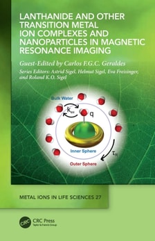
Since the development of the first clinical MRI contrast agent, Gd(DTPA) (or Magnevist(R)) in the early 1980s, another three linear and three macrocyclic (eg.
Clinical applications include: detecting pre-cancerous and cancerous tissue states; characterizing cell and tissue properties for identifying disease; and assessing the presence and concentration of biochemicals for diagnostic purposesPart of the McGraw-Hill Biophotonics Series