When compiling the present atlas our aim has been to provide the practis- ing radiotherapist with a handbook which would help him to plan the radiation therapy of tumours of individual organs.
Over the last few decades, angiography has devel- careful analysis of his angiographic findings and a oped enormously and an extensive literature has very thorough description of vascular anatomy been published on the subject.
This atlas is a selection of roentgenograms of patients who visited the radiology departments at the University Hospital in Leiden between 1970 and 1978, the Free University Hospital in Amsterdam in 1979, and the radiology department at the Indiana University Medical School in Indianapolis in 1977.
Complex disorders of the carbohydrate metabolism and associated complications cause many abnormalities detectable by radiography in the bones and joints.
In 1977 a Philips Tomoscan 200, second generation, whole body CT scanner was installed at the Department of Radiodiagnosis of the University Hospital of Utrecht (The Netherlands) and its new possibilities concerning the measurements of bone mineral content (BMC) had been considered.
The liver blood flow disorders are usually described under the term 'portal hypertension', which is a well- known syndrome since it has been widely studied for years from a clinical, radiological and therapeutic point of view.
I hope this book, which covers the Equipment section of With the help of the Superintendent find out which quality the DCR and HDCR syllabuses, will be of help not only assurance tests are carried out on the equipment and ask to those students preparing for these examinations, but for permission to participate in the procedures.
This book is designed as a definitive report on current capabilities of ultrasound imaging and Doppler evaluation of the cerebral circulation, both extracranial and intracranial.
Computed tomography is presently reaching maturity with its high-resolution reconstruction programs, as a result of which conventional tomography has definitely been surpassed.
Safety and Efficacy of Radiopharmaceuticals was established as a very important and comprehensive subject at the First Europe- an Symposium on Radiopharmacy and Radiopharmaceuticals in Denmark in 1983.
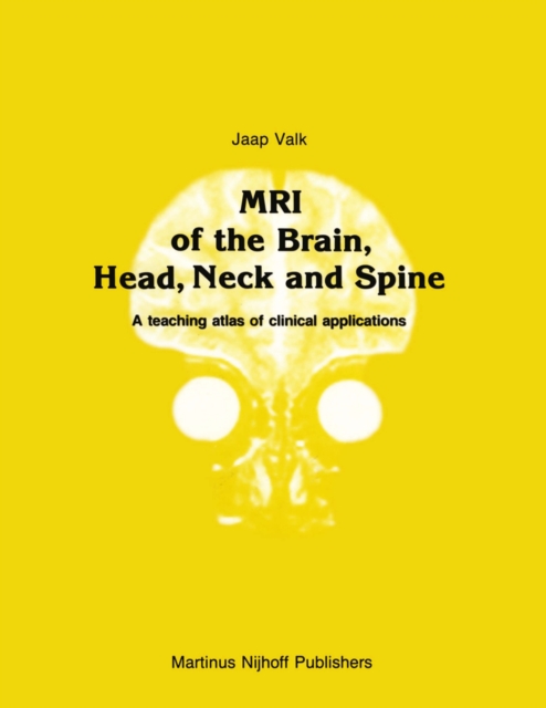
With the growing number of MR installations, clinicians and radiologist are being confronted more and more with visual information they do not feel as confident with as with the more 'mono-form' infor- mation of conventional radiographs, CT and US.
I am particularly pleased to be able to write the introduction to this book that resulted from a collaborative effort by the Radiology De- partment, under the auspices of the Boerhaave Committee for Postgraduate Medical Education of the Faculty of Medicine, at the University of Leiden and the Department of Medical Imaging and Radiological Sciences at Van- derbilt University.
Publication of this book has been stimulated by 15 performed and duodenal abnormalities thus not years experience with contrast fluid irrigation visualised, occurred frequently.
This monograph represents an attempt to collect the methods developed within the past decade in which echocardiography was being used to guide or to assess the results of some cardiovascular interventions or to overcome the imaging limitations of transthoracic approaches.
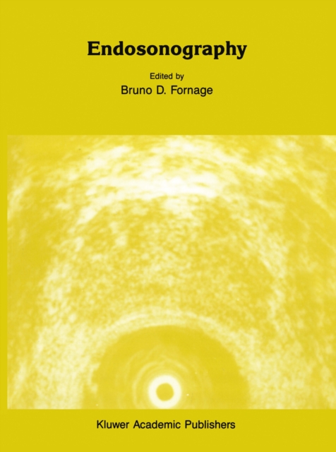
Following the development of gray-scale imaging, real-time scanning, Doppler examination, and high-frequency sonography, endosonography is one of the latest major breakthroughs in the history of diagnostic ultrasound.
In the last few years ways of thinking in psychiatry have undergone considerable change thanks to advances in the fields of morphology and plasticity of the CNS, particularly with regard to schizophrenic and mood disorders.
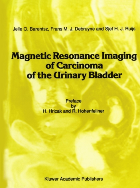
Carcinoma of the urinary bladder is a common (in the USA it is the fifth most common form of cancer in males and tenth most common form of cancer in females) malignan- cy and one in which noninvasive staging by imaging plays such an important role.
During the past decade Positron Emission Tomography (PET) has turned from a highly sophisticated tool developed for basic research in neurology and cardiology into an advanced nuclear medicine imaging technique ready for routine use.
Radiophannaceuticals labeled with short-lived radionuclides are utilized to unravel biochemical processes, and to diagnosis and treat diseases of the living body are-developed through extensive evaluation in ~iological models.
With strong numerical and computational focus, this book serves as an essential resource on the methods for functional neuroimaging analysis, diffusion weighted image analysis, and longitudinal VBM analysis.
This book is the product of a unique collaboration by experts from leading international, regional and national agencies and professional organizations discussing on the current 'hot' issue on the judicious use and safety of radiation in radiology.
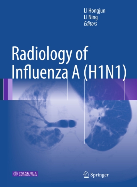
A novel strain of influenza A (H1N1) virus emerged and an outbreak of influenza A (H1N1) occurred worldwide in 2009 and 2010, posing a severe threat to the health of human beings.
This volume will cover what is known regarding the biology and treatment of CNS metastasis, including novel chapters such as the future of targeted therapies.
Since the early 20th century, medical imaging has been dominated by monochrome imaging modalities such as x-ray, computed tomography, ultrasound, and magnetic resonance imaging.