The 17th volume of the "e;Advances in Neurosurgery"e; contains a selection of the scientific reports of the 39th annual meeting of the German Society for Neurosurgery, which was held in Cologne from May 8-11, 1988.
This 16th volume of Advances in Neurosurgery contains a selection of pa- pers presented at the 38th Annual Meeting of the German Society of Neurosurgery, held in Munster on 3-6 May 1987.
Die spontanen intrazerebralen Hämatome sind die zweithäufigste zerebro-vaskuläre Erkrankung nach den ischämischen Hirninfarkten und gehören daher seit langem in Neurologie, Neurochirurgie und Neuropathologie zu den traditionellen Forschungsgebieten.
The aim of the book is to describe the current approach to meningiomas on the basis of experience gained in the fields of histopathology, biology, radiology and surgery.
Based on the principles of functional vascular anatomy and endovascular treatment described in the first three volumes of Surgical Neuroangiography, Volumes 4 and 5 complete the series that takes a revolutionary approach in endovascular neurosurgery.
In Neuroorthopädie Band 4 werden die Erkrankungen des zervikookzipitalen Übergangs mit Beteiligung des Nervensystems in Diagnostik und Therapie, zumal deren operative Behandlung, diskutiert.
Embolization has been performed in many European countries and in North America for over 20 years and is now beginning to gain accep- tance in other countries.
In this age when we are witnessing a veritable explosion in new modalities in diagnos- tic imaging we continue to have a great need for detailed studies of the vascularity of the brain in patients who have all types of cerebral vascular disease.
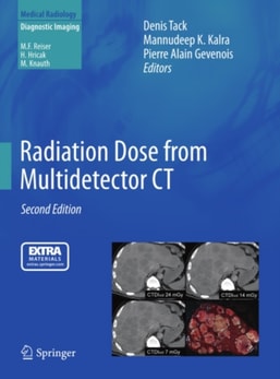
The introduction of multidetector spiral CT into clinical practice is without any doubt one of the most important technical developments in the field of computed tomography in general, and spiral CT in particular, in recent years.
The numerous ways in which man and animals are affected by their physical environment, and the inborn and adaptive responses to change in the "e;milieu exterieur"e; have fascinated curious minds since the earliest days of recorded history.
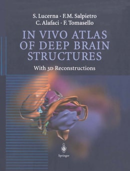
In the first half of the twentieth century, the study of neuroanatomy was essentiallybased on the observations made by scientists on brain cadavers fixed with standard techniques.
In the past two decades much has been published on whiplash injury, yet both the confusion regarding the condition and the medicolegal discussion surrounding it have increased.
This book is based on contributions presented at the 1st World Congress on Gallium-68 and Peptide Receptor Radionuclide Therapy, which examined recent developments in theranostics - the emerging field of molecular targeting of vectors that can be used for both diagnosis and therapy, when modified accordingly.