Magnetic Resonance Angiography: Principles and Applications is a comprehensive text covering magnetic resonance angiography (MRA) in current clinical use.
Functional magnetic resonance imaging (fMRI) measures quick, tiny metabolic changes that take place in the brain, providing the most sensitive method currently available for identifying, investigating, and monitoring brain tumors, stroke, and chronic disorders of the nervous system like multiple sclerosis, and brain abnormalities related to dementia or seizures.
This volume acquaints the non-neuropathologist with the advantages of clinical-radiologic-pathologic correlation in neuropathology specimens, particularly in the intra-operative consultation.
Evidence-Based Imaging in Pediatrics: Optimizing Imaging in Pediatric Patient Care presents a user friendly guide to the evidence-based science and the merit defining the appropriate use of medical imaging in infants and children.
This compendium of cardiac CT angiographic cases is a sampling of the breadth of real-world cases encountered in a busy practice with examples of pathology and normal variants.
Medical Imaging Informatics provides an overview of this growing discipline, which stems from an intersection of biomedical informatics, medical imaging, computer science and medicine.
Functional Neuroradiology: Principles and Clinical Applications, is a follow-up to Faro and Mohamed's groundbreaking work, Functional (BOLD)MRI: Basic Principles and Clinical Applications.
Drug development today needs to balance agility, speed, and risk in defining probability of success for molecules, mechanisms, and therapeutic concepts.
This encyclopedia examines more than 125 of the most important and commonly performed medical tests, providing readers with information about how and why they are performed and how each test contributes to monitoring health and diagnosing and treating medical conditions.
With every chapter revised and updated, Physics for Diagnostic Radiology, Third Edition continues to emphasise the importance of physics education as a critical component of radiology training.
Despite a number of books on biophotonics imaging for medical diagnostics and therapy, the field still lacks a comprehensive imaging book that describes state-of-the-art biophotonics imaging approaches intensively developed in recent years.
While there are many excellent texts focused on clinical medical imaging, there are few books that approach in vivo imaging technologies from the perspective of a scientist or physician-scientist using, or interested in using, these techniques in research.
Cardiovascular and Neurovascular Imaging: Physics and Technology explains the underlying physical and technical principles behind a range of cardiovascular and neurovascular imaging modalities, including radiography, nuclear medicine, ultrasound, and magnetic resonance imaging (MRI).
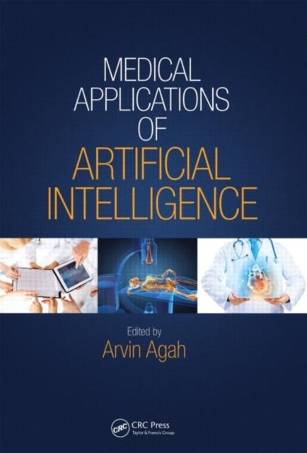
Enhanced, more reliable, and better understood than in the past, artificial intelligence (AI) systems can make providing healthcare more accurate, affordable, accessible, consistent, and efficient.
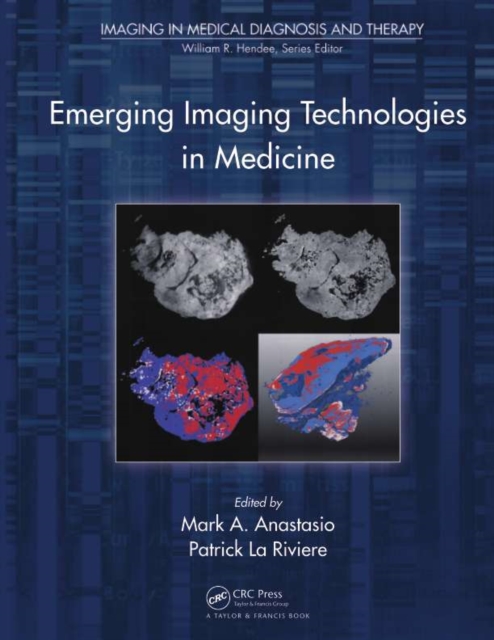
From the discovery of x-rays in 1895 through the emergence of computed tomography (CT) in the 1970s and magnetic resonance imaging (MRI) in the 1980s, non-invasive imaging has revolutionized the practice of medicine.
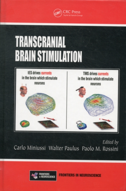
Since the discovery of transcranial magnetic stimulation (TMS) and transcranial electrical stimulation (tES), these non-invasive brain stimulation (NIBS) techniques have been used to investigate the state of cortical excitability, and the excitability of the cortico-cortical and corticospinal pathways.
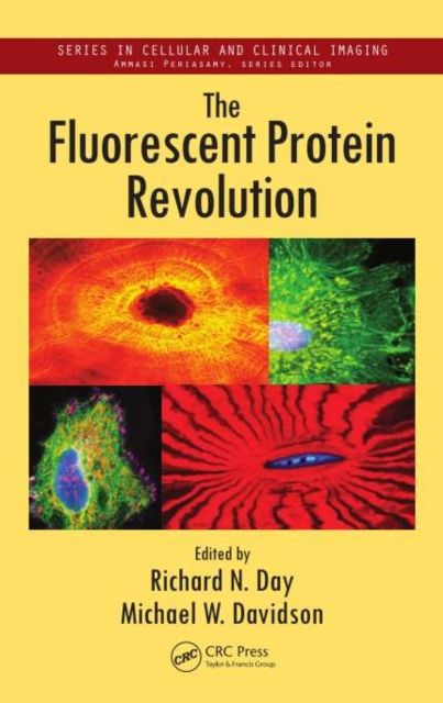
Advances in fluorescent proteins, live-cell imaging, and superresolution instrumentation have ushered in a new era of investigations in cell biology, medicine, and physiology.
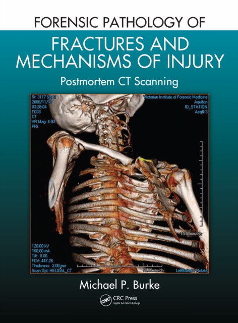
Practitioners of forensic medicine have various tools at their disposal to determine cause of death, and today's computed tomography (CT) can provide valuable clues if images are interpreted properly.
An innovative, three-dimensional x-ray imaging technique that enhances projection radiography by adding depth resolution, Tomosynthesis Imaging explores tomosynthesis, an emerging limited-angle tomographic imaging technology that is being considered for use in a range of clinical applications, and is currently being used for breast cancer screening
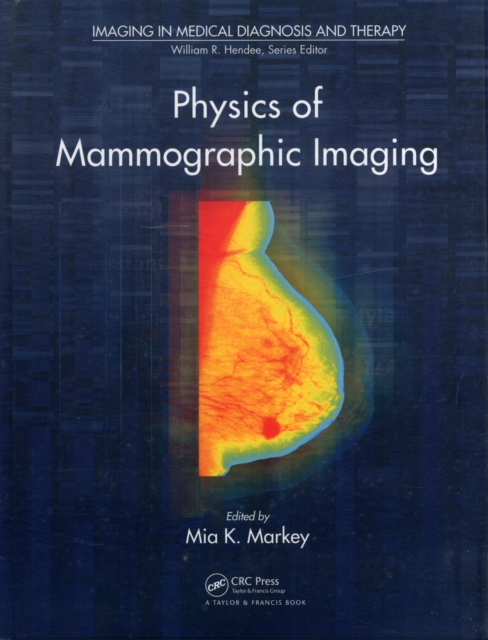
Due to the increasing number of digital mammograms and the advent of new kinds of three-dimensional x-ray and other forms of medical imaging, mammography is undergoing a dramatic change.
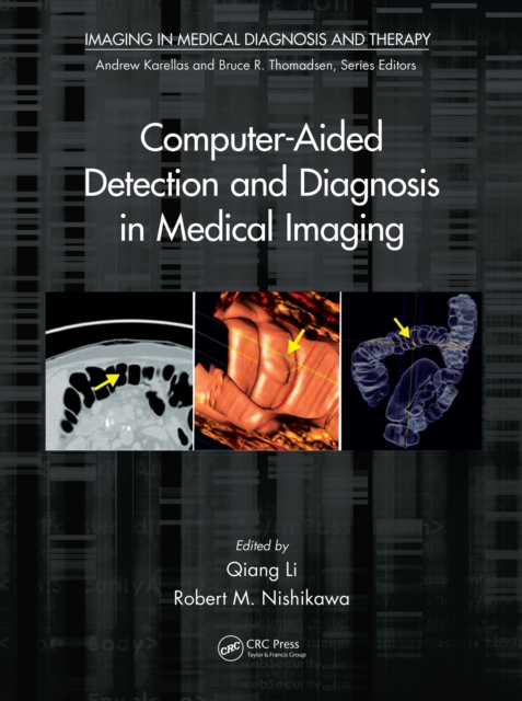
Improve the Accurate Detection and Diagnosis of Cancer and Other DiseasesDespite the expansion of the CAD field in recent decades, there is currently no single book dedicated to the development and use of CAD systems.
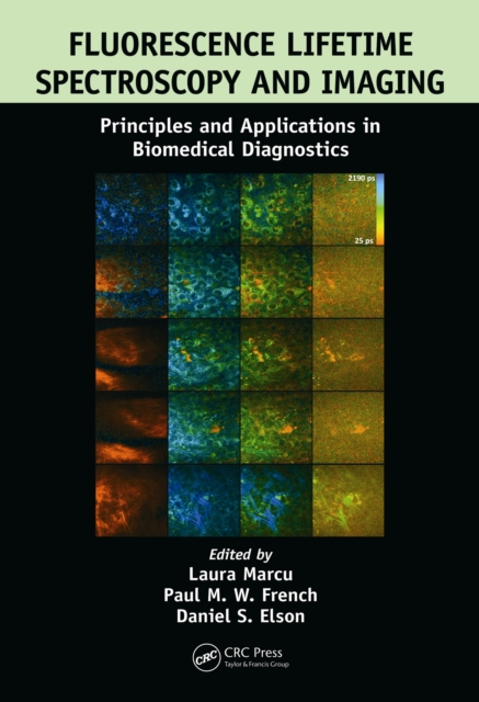
During the past two decades, there has been an increasing appreciation of the significant value that lifetime-based techniques can add to biomedical studies and applications of fluorescence.
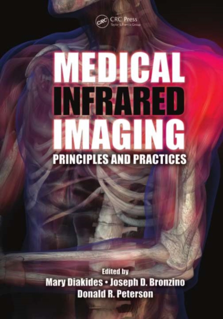
The evolution of technological advances in infrared sensor technology, image processing, "e;smart"e; algorithms, knowledge-based databases, and their overall system integration has resulted in new methods of research and use in medical infrared imaging.
The First Book on CRS MicroscopyCompared to conventional Raman microscopy, coherent Raman scattering (CRS) allows label-free imaging of living cells and tissues at video rate by enhancing the weak Raman signal through nonlinear excitation.
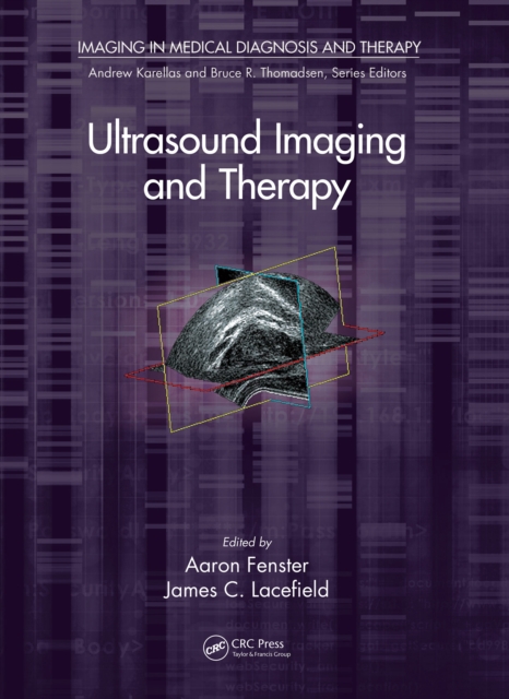
Up-to-Date Details on Using Ultrasound Imaging to Help Diagnose Various DiseasesDue to improvements in image quality and the reduced cost of advanced features, ultrasound imaging is playing a greater role in the diagnosis and image-guided intervention of a wide range of diseases.
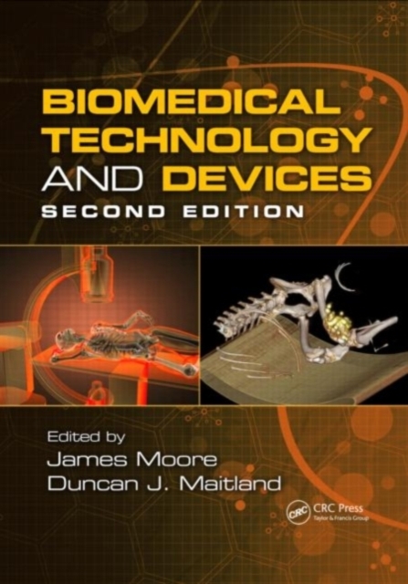
Fully revised since its original publication, this reference illuminates the equipment, devices, and techniques used in modern medicine to diagnose, treat, and monitor human illnesses.
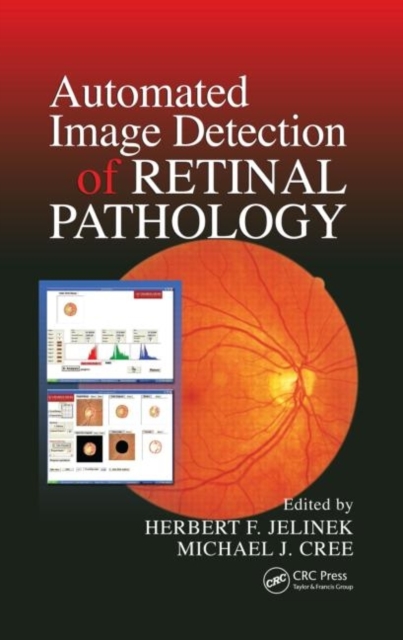
Discusses the Effect of Automated Assessment Programs on Health Care ProvisionDiabetes is approaching pandemic numbers, and as an associated complication, diabetic retinopathy is also on the rise.
Of all children reported to child protective services for suspected maltreatment in any form, the percentage of substantiated cases of actual physical abuse is quite small.
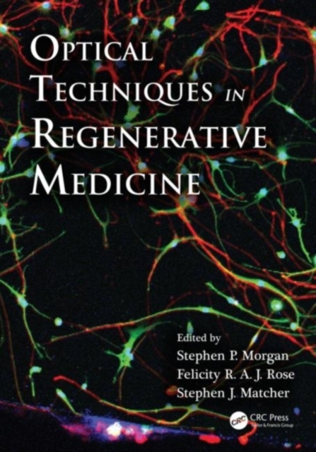
In regenerative medicine, tissue engineers largely rely on destructive and time-consuming techniques that do not allow in situ and spatial monitoring of tissue growth.
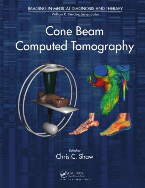
Conventional computed tomography (CT) techniques employ a narrow array of x-ray detectors and a fan-shaped x-ray beam to rotate around the patient to produce images of thin sections of the patient.
In the medical imaging field, clinicians and researchers are increasingly moving from the qualitative assessment of printed images to the quantitative evaluation of digital images since the quantitative techniques often improve diagnostic accuracy and complement clinical assessments by providing objective criteria.
Details on specific imaging modalities for different cellular and tissue engineering applications are scattered throughout articles and chapters in the literature.
Second-harmonic generation (SHG) microscopy has shown great promise for imaging live cells and tissues, with applications in basic science, medical research, and tissue engineering.
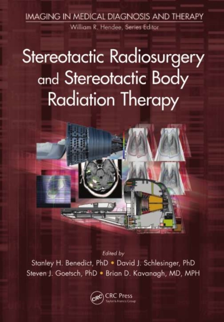
Written by internationally known experts in the field, Stereotactic Radiosurgery and Stereotactic Body Radiation Therapy examines one of the fastest-developing subspecialties within radiation oncology.
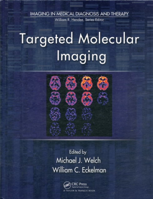
Targeted Molecular Imaging covers the development of novel diagnostic approaches that use an imaging probe and agent to noninvasively visualize cellular processes in normal and disease states.
Biomedical Applications of Magnetic Particles discusses fundamental magnetic nanoparticle physics and chemistry and explores important biomedical applications and future challenges.
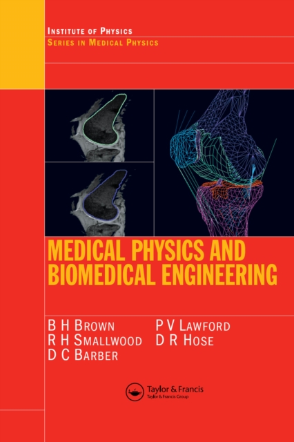
Medical Physics and Biomedical Engineering provides broad coverage appropriate for senior undergraduates and graduates in medical physics and biomedical engineering.
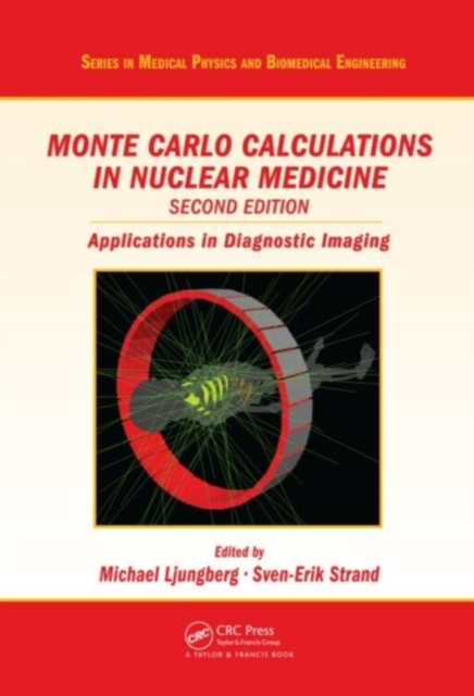
From first principles to current computer applications, Monte Carlo Calculations in Nuclear Medicine, Second Edition: Applications in Diagnostic Imaging covers the applications of Monte Carlo calculations in nuclear medicine and critically reviews them from a diagnostic perspective.