This comprehensive volume demonstrates the broad scope of uses for data mining and includes detailed strategies and methodologies for analyzing data from biomedical images, signals, and systems.
Images from CT, MRI, PET, and other medical instrumentation have become central to the radiotherapy process in the past two decades, thus requiring medical physicists, clinicians, dosimetrists, radiation therapists, and trainees to integrate and segment these images efficiently and accurately in a clinical environment.
Reflecting the increased importance of the collaborations between radiation oncology and informatics professionals, Informatics in Radiation Oncology discusses the benefits of applying informatics principles to the processes within radiotherapy.
As the third volume of The Biomedical Engineering Handbook, Fourth Edition, this book covers broad areas such as biosignal processing, medical imaging, infrared imaging, and medical informatics.
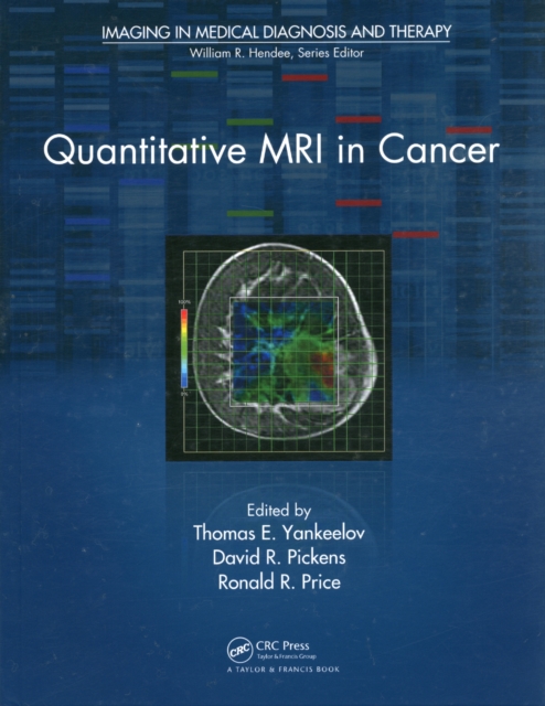
Propelling quantitative MRI techniques from bench to bedside, Quantitative MRI in Cancer presents a range of quantitative MRI methods for assessing tumor biology.
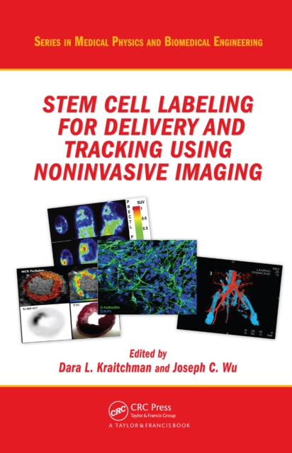
Stem Cell Labeling for Delivery and Tracking Using Noninvasive Imaging provides a comprehensive overview of cell therapy imaging, ranging from the basic biology of cell therapeutic choices to the preclinical and clinical applications of cell therapy.
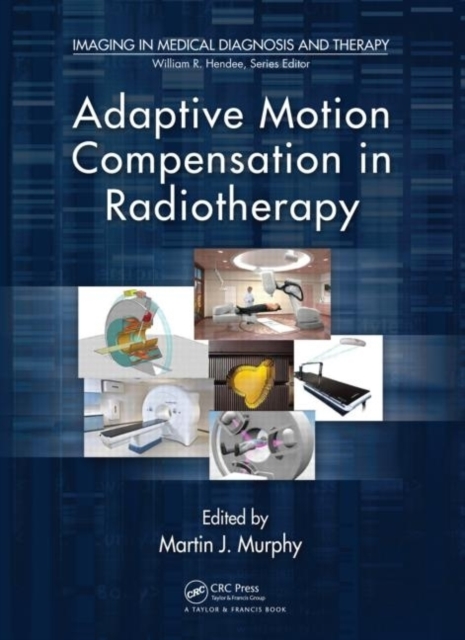
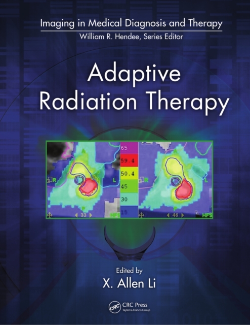
Modern medical imaging and radiation therapy technologies are so complex and computer driven that it is difficult for physicians and technologists to know exactly what is happening at the point-of-care.
Written by an interdisciplinary team of medical doctors, computer scientists, physicists, engineers, and mathematicians, Correction Techniques in Emission Tomography presents various correction methods used in emission tomography to generate and enhance images.
Uses the FPT to Solve the Quantification Problem in MRSAn invaluable tool in non-invasive clinical oncology diagnosticsAddressing the critical need in clinical oncology for robust and stable signal processing in magnetic resonance spectroscopy (MRS), Signal Processing in Magnetic Resonance Spectroscopy with Biomedical Applications explores cutting-
Endometriosis affects women in the reproductive years, is associated with pelvic pain and infertility, and - although not life threatening - can seriously impair health, with huge economic and social consequences.
Since publication of the First Edition, advances in the diagnosis and prevention and treatment strategies of the vulnerable plaque have necessitated this greatly expanded second edition.
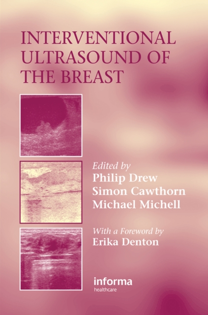
Over the past decade interventional radiology techniques have replaced surgical and clinically guided diagnostic procedures such that the vast majority of patients have a non surgical diagnosis of a breast problem.
The last 20 years have brought unprecedented new knowledge to our understanding of Alzheimer's disease (AD) and for the first time, approved symptomatic treatments.
This book provides an in-depth review of knowledge of the corpus callosum, called white matter or terra incognita, with emphasis on anatomical, embryological, diagnostics, and surgical features.
This book offers a comprehensive exploration of brain tumors, beginning with a foundational understanding of their pathophysiology and extending through the latest advancements in diagnosis and treatment.
This book describes the role of radioactivity in the living world and highlights the many aspects of the impact of ionizing radiation on biological systems.
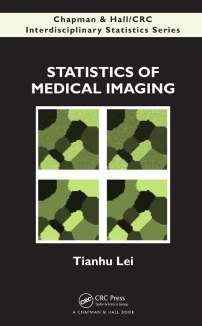
More work is being done in the statistical aspects of medical imaging, and this book fills the gap to provide a unified framework of study by presenting a complete look at medical imaging and statistics - from the statistical aspects of imaging technology to the statistical analysis of images.
Shaped by Quantum Theory, Technology, and the Genomics RevolutionThe integration of photonics, electronics, biomaterials, and nanotechnology holds great promise for the future of medicine.
Shaped by Quantum Theory, Technology, and the Genomics RevolutionThe integration of photonics, electronics, biomaterials, and nanotechnology holds great promise for the future of medicine.
Bringing the long tradition of radiologic pathologic correlation to forensic radiology and autopsy, this volume provides readers with a technical and interpretive foundation for applying modern cross-sectional imaging to forensic autopsy.
Medical and industrial imaging methods have come to be recognized as powerful tools for documentation and data collection in many nontraditional settings.
Written by skilled specialists in the field of interventional pulmonology, the new Second Edition thoroughly explores the latest advancements, newest therapies, and diagnostic techniques in interventional pulmonary medicine.
Detecting Signals at the Single Molecule Level: Pioneering Achievements in MicroscopyRecent advances have led to such remarkable improvements in fluorescence lifetime imaging microscopy's (FLIM) capacity for contrast and sensitivity that researchers can now employ it to detect signals at the single molecule level.
Combining a professional development course on diagnostic endoscopy from SPIE (the international society advancing light-based research) and the authors' graduate course on biomedical optics, this work is written for researchers in medical optics and biomedical engineering as well as graduate medical optics students.
To improve efficiency and reduce administrative costs, healthcare providers, insurance companies, and governments are increasingly using integrated electronic health record (EHR) and picture archiving and communication systems (PACS) to manage patients' medical information.
The benchmark first edition of Forensic Radiology, published in 1998, was a milestone in the forensic community a "e; a bestseller throughout the world and a standard reference for practitioners and educators alike.
The tools of nanodiagnostics, nanotherapy, and nanorobotics are expected to revolutionize the future of medicine, leading to presymptomatic diagnosis of disease, highly effective targeted treatment therapy, and minimum side effects.
In considering ways that physics has helped advance biology and medicine, what typically comes to mind are the various tools used by researchers and clinicians.
Discover the Applicability, Benefits, and Potential of New Technologies As advances in algorithms and computer technology have bolstered the digital signal processing capabilities of real-time sonar, radar, and non-invasive medical diagnostics systems, cutting-edge military and defense research has established conceptual similarities in these areas.
With over 100 illustrations, Volume 1 addresses the core disciplines of pharmaceutics (absorption, PK, excipients, tablet dosage forms, and packaging), and explores the challenges and paradigms of pharmaceutics.
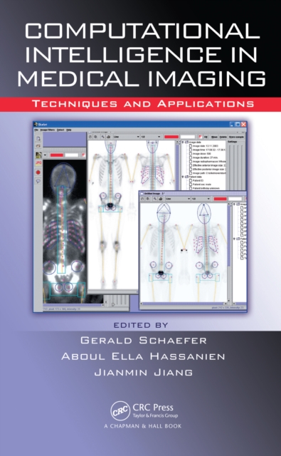
CI Techniques & Algorithms for a Variety of Medical Imaging SituationsDocuments recent advances and stimulates further researchA compilation of the latest trends in the field, Computational Intelligence in Medical Imaging: Techniques and Applications explores how intelligent computing can bring enormous benefit to existing technology in medical
Photoacoustics promises to revolutionize medical imaging and may well make as dramatic a contribution to modern medicine as the discovery of the x-ray itself once did.
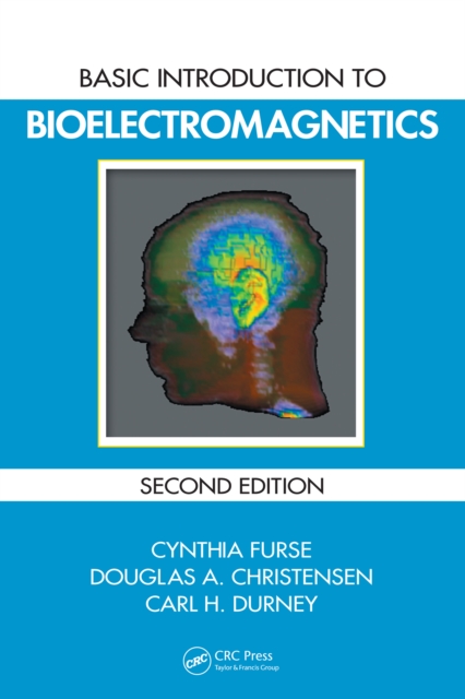
Although classical electromagnetic (EM) field theory is typically embedded in vector calculus and differential equations, many of the basic concepts and characteristics can be understood with precursory mathematical knowledge.
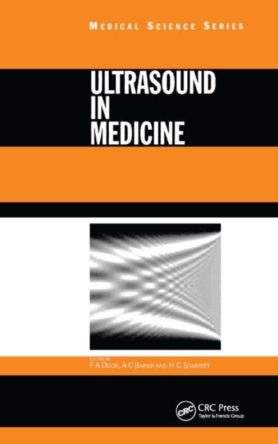
Ultrasound in Medicine is a broad-ranging study of medical ultrasound, including ultrasound propagation, interaction with tissue, and innovations in the application of ultrasound in medicine.
The Ischemic Penumbra presents the current status of concepts and research on this topic and identifies the latest methods for clinicians to quickly and efficiently recognize viable cerebral tissue for enhanced stroke management.
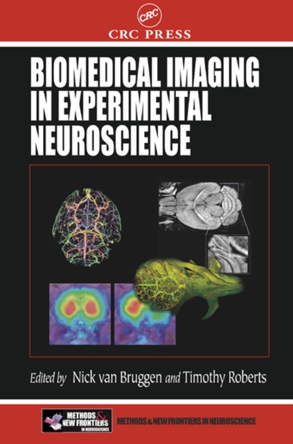
With the emergence of genetically manipulated laboratory mice as one of the most powerful tools for neuroscientists, imaging techniques capable of providing anatomical and functional information of small animals have become extremely important.
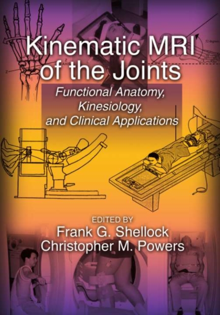
Kinematic MRI refers to imaging a joint through a range of motion to examine the interactions between the soft tissue and osseous anatomy that comprise the joint.