The objective of this Atlas is to contribute to the knowledge of morphological findings of the main cardiovascular diseases among the various specialists who deal with people affected of heart diseases: cardiologists and cardiovascular surgeons, clinical and forensic pathologists, coroners, sports medicine doctors.
This book is designed to meet the needs of radiologists and radiographers by clearly depicting the anatomy that is generally visible on imaging studies.
This is the first atlas to depict in high-resolution images the fine structure of the spinal canal, the nervous plexuses, and the peripheral nerves in relation to clinical practice.
The book provides a comprehensive description of the basic ultrasound principles, normal anatomy of the lower limb muscles and classification of muscle strain injuries.
Teaching Anatomy: A Practical Guide is the first book designed to provide highly practical advice to both novice and experienced gross anatomy teachers.
This book, featuring more than 180 high spatial resolution images obtained with state-of-the-art MDCT and MRI scanners, depicts in superb detail the anatomy of the temporal bone, recognized to be one of the most complex anatomic areas.
The comprehensive approach to anatomy is a new attempt to understand the organization of anatomical structures instead of only memorizing details, which is both time-consuming and prone to error.
This atlas illustrates the anatomical structures of the upper basal cisterns, their topography and relationship to other intra- and extradural structures.
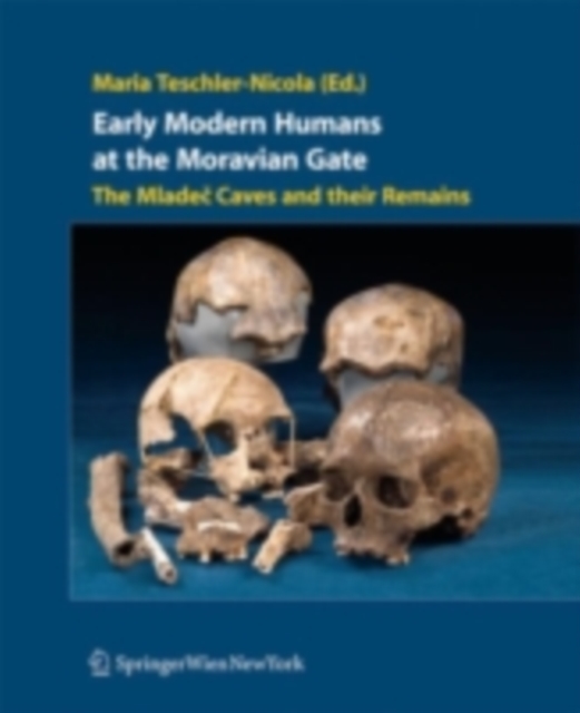
This beautifully illustrated monograph addresses - for the first time - the complete assemblage of the Mladec caves finds, including the human cranial, post cranial, teeth and jaw fragments of several individuals as well as faunal remains and archaeological objects.
Color Atlas of Human Anatomy, Volume 2: Internal OrgansFor over 45 years, the three-volume Color Atlas of Human Anatomy has provided readers with a compact review of the human body and its structures.
Color Atlas of Human Anatomy, Volume 1: Locomotor SystemFor over 45 years, the three-volume Color Atlas of Human Anatomy has provided readers with a compact review of the human body and its structures.
A proven, didactic guide to normal ultrasound anatomy ideal for the beginning sonographerColor Atlas of Ultrasound Anatomy, Second Edition presents a systematic, step-by-step introduction to normal sectional anatomy of the abdominal and pelvic organs and thyroid gland, essential for recognizing the anatomic landmarks and variations seen on ultrasound.
In this easily accessible pocket atlas, two expert radiologists present the normal radiographic anatomy readers need in order to interpret conventional diagnostic radiographs.
Whatever are your beliefs, background, education, political views or interests, one thing is sure: this book will engage you, teach you something new, and more importantly make you to re-think deeply about critical aspects of your daily-life, including sex, love, food, physical activities, diseases, work and stress, and how you see and deal with other people, other animals, and the planet in general.
Das Taschenwörterbuch erklärt kurz und knapp die wichtigsten 2200 medizinischen Fachbegriffe hinsichtlich ihrer sprachlichen Herkunft, teilweise auch geschichtlicher u.
This book brings together current advances in high-technology visualisation and the age-old but science-adapted practice of drawing for improved observation in medical education and surgical planning and practice.
This book presents a detailed examination of the current state of knowledge in the field of paleoneurology in the main amniote groups (reptiles, birds and mammals), and advances resulting from new non-invasive technologies.
This image-rich book explores the practice as well as the theory of visual representation and presents us with the importance of designing appropriate images for communication to specific target audiences.